Download
1 / 6
60 likes | 63 Views
Chronic venous insufficiency (CVI) may lead to leg ulceration (CVI stage C6) (Nicolaides<br>A.N. et al., 2000). Some authors published that genetic risk factors are observed more frequently in<br>patients with CVI stage C6.

E N D
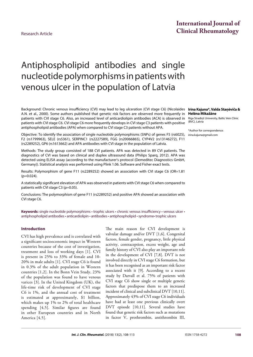
International Journal of Clinical Rheumatology Research Article Antiphospholipid antibodies and single nucleotide polymorphisms in patients with venous ulcer in the population of Latvia Background: Chronic venous insufficiency (CVI) may lead to leg ulceration (CVI stage C6) (Nicolaides A.N. et al., 2000). Some authors published that genetic risk factors are observed more frequently in patients with CVI stage C6. Also, an increased level of anticardiolipin antibodies (ACA) is observed in patients with CVI stage C6. CVI stage C6 more frequently develops in CVI stage C3 patients with positive antiphospholipid antibodies (APA) when compared to CVI stage C3 patients without APA. Irina Kajuna*, Valda Staņēviča & Helēna Mikažāne Rīga Stradiņš University, Baltic Vein Clinic (BVC), Latvia *Author for correspondence: irina.kajuna@gmail.com Objective: To identify the association of single nucleotide polymorphisms (SNPs) of genes F5 (rs6025), F2 (rs1799963), SELE (rs5361), SERPINC1 (rs2227589), FGG (rs20066865), CYP4V2 (rs13146272), F11 (rs2289252), GP6 (rs1613662) and APA antibodies with CVI stage in the population of Latvia. Methods: The study group consisted of 188 CVI patients. APA was detected in 89 CVI patients. The diagnostics of CVI was based on clinical and duplex ultrasound data (Philips Sparq, 2012). APA was detected using ELISA assay (according to the manufacturer’s protocol (Demeditec Diagnostics GmbH, Germany)). Statistical analysis was performed using Plink 1.06. Software and Fisher exact tests. Results: Polymorphism of gene F11 (rs2289252) showed an association with CVI stage C6 (OR=1.81 (p=0.024). A statistically significant elevation of APA was observed in patients with CVI stage C6 when compared to patients with CVI stage C3 (p=0.05). Conclusions: The polymorphism of gene F11 (rs2289252) and positive APA showed an association with CVI stage C6. Keywords: single nucleotide polymorphisms • trophic ulcers • chronic venous insufficiency • venous ulcer • antiphospholipid antibodies • anticardiolipin • antibodies • antiphospholipid • syndrome• trophic ulcers Introduction The main reason for CVI development is valvular damage and/or DVT [1,6]. Congenital factors, female gender, pregnancy, little physical activity, contraception, excess weight, age and family history of CVI also play an important role in the development of CVI [7,8]. DVT is not involved directly in CVI stage C6 formation, but it has been recognised as an important risk factor associated with it [9]. According to a recent study by Darvall et al. 75% of patients with CVI stage C6 show single or multiple genetic factors that predispose them to an increased incident of clinical and subclinical DVT [10,11]. Approximately 43% of CVI stage C6 individuals have had at least one previous clinically overt DVT episode [10,11]. Several studies have found that genetic risk factors such as mutations in factor V, prothrombin, antithrombin III, CVI has high prevalence and is correlated with a significant socioeconomic impact in Western countries because of the cost of investigation, treatment and loss of working days [1]. CVI is present in 25% to 33% of female and 10- 20% in male adults [1]. CVI stage C6 is found in 0.3% of the adult population in Western countries [1,2]. In the Bonn Vein Study, 23% of the population was found to have venous varices [3]. In the United Kingdom (UK), the life-time risk of development of CVI stage C6 is 1%, and the annual cost of treatment is estimated at approximately, $1 billion, which makes up 1% to 2% of total healthcare spending [4,5]. Similar figures are found in other European countries and in North America [4,5]. 108 Int. J. Clin. Rheumatol. (2018) 13(2), 108-113 ISSN 1758-4272
Research Article Kajuna, Staņēviča, Mikažāne protein C and S genes is found more frequently in patients with CVI stage C6 (44–75%) [12]. In the general population, the prothrombin 20210A variant is found in 2% of individuals. In the patients with DVT the prothrombin 20210A variants is found in approximately 6% of individuals and in the patients with CVI stage C6 up to 4% of individuals [10,13-15]. Factor V Leiden the mutant allele is found at a frequency of approximately 5% in Caucasian populations; the prevalence of this risk allele in patients with recognised DVT is as high as 30%, whereas in the group of patients with CVI stage C6 it ranges from between 8 and 36%, depending on the estimated population size [2,10]. It is important to characterize populations using molecular tools. The applications of molecular genetics have many important advantages [16], hence determination of gene polymorphism is important in different populations [17-19]. Some studies have shown that CVI stage C6 develops more frequently in patients with CVI stage C3 with positive APA [20,21]. The pathogenic mechanism of venous ulcer formation involves tissue oedema, hyperpigmentation, skin atrophy before ulcer development [20- 22]. Microlymphangiopathy, lymphatic vessel dilatation and capillary dilatation, elongation and microthrombotic occlusion are found at a microvascular level [20-22]. APA is a heterogeneous group of autoantibodies directed against phospholipid-binding plasma proteins (most commonly beta-2-glycoprotein (B2GP)) [23]. APA are also produced against proteins participating in the coagulation process — prothrombin, protein C, protein S and others [24]. The causes of APA formation include infections and drugs [23]. Increased levels of ACA during infection are usually transient, and IgM antibodies are elevated more frequently than IgG [23]. Materials and Methods The control group consisted of 188 individuals selected from the Genome database of the Latvian population (LGDB). Individuals with diagnoses I11 to I99 (according to ICD10) were not included in the control group. The case and control group were matched for sex, age and BMI. The control group characteristics: for CVI C3 patients control group 28 men and 98 women, average age 46.3 ± 13.1 years, BMI 27.2 ± 4.6; for CVI C6 patients control group 14 men and 48 women, average age 61.9 ± 11.9 years, BMI 30.2 ± 5.7. The adjustment for sex, age, BMI was performed to exclude the possible influence of these factors on association results, as the groups were closely matched, however, not identical and it was not possible to ensure matching of the groups in the stratification analysis. DNA samples from white blood cells (WBC) were diluted using Freedom Evo robotic workstation (Tecan, Männedorf, Switzerland) with disposable filter tips into 372 wells of 380- well PCR plates (28 ng per well), eight positions in the plate was filled with Millipore (Millipore, Bedford, USA) H2O for negative control (each for one TaqMan SNP genotyping probe). Genotyping was performed using Biosystems TaqMan SNP Genotyping Assay according to the manufacturer’s protocol in ViiA™ 7 real-time PCR system. We used SNP genotyping assays: C_11975332_10 for rs5361, C_11503414_10 for rs2066865, C_16180170_20 for rs2227589, C_8717873_10 for rs1613662, C_25990131_20 for rs13146272, C_3230038_10 for rs2289252, C_11975250_10 for rs6025 and C_8726802_20 for rs1799963 (Applied Biosystems, USA). Genotypes were assigned using AutoCaller 1.1 (Applied Biosystems, USA) software. Statistical analysis was performed using Plink 1.06 software. All genotyped polymorphisms corresponded to the Hardy-Weinberg equation and the efficacy of first time SNP genotyping was 98.8%. The additive inheritance model was used in logistic regression analysis and adjusted for gender, age and body mass index. Antiphospholipid IgM and IgG, and anticardiolipin (ACA) IgM and IgG antibodies were detected using the ELISA assay (according to the manufacturer’s protocol (Demeditec Diagnostics GmbH, Germany)). Statistical analysis was performed using Plink 1.06 software; Fisher’s exact tests were used. Results The study group consisted of 188 patients with CVI stage C3–C6 (including 62 patients with CVI stage C6). AFA were detected in 89 CVI patients (fifty-three (53) patients had CVI stage C3 and 36 patients had CVI stage C6). The inclusion criteria for the study group: patient is 18 years of age or above, the patient is not mentally retarded, the patient is diagnosed with one of the following diseases —CVI, trophic ulcer, optional criterion — family history of DVT. The diagnostics were based on clinical and duplex ultrasound data (Philips Sparq, 2012). In the study, we used data from patients diagnosed with CVI and analysed their association with 8 genes SNP from extracted DNA. We analysed the association of APA with the CVI stage. 109 Int. J. Clin. Rheumatol. (2018) 13(2)
Antiphospholipid antibodies and single nucleotide polymorphisms in patients with Research Article venous ulcer in the population of Latvia The phenotypic characteristic of the study groups are given in Tables 1 and 2. Commonly the patients of the study group were middle-aged women with increased BMI. When comparing the frequency of risk alleles in patient and control groups, we found a trend for the risk allele of the analysed FGG (rs20066865) gene to be found more frequently in CVI C6 (p=0.019) patients Table 3. Table 4 shows the association of the analysed SNPs in the CVI C6 group. Polymorphism of gene F11 (rs2289252) showed a significant association with CVI stage C6 (OR=1.81 (p=0.024)). Polymorphism of genes F5 (rs6025), F2 (rs1799963), SERPINC1 (rs2227589), CYP4V2 (rs13146272), FGG (rs20066865) and GP6 (rs1613662) did not show a significant relationship with CVI C6 Table 4. AFA were detected in 89 CVI patients. From the 53 CVI stage C3 patients, 8 (15%) were men and 45 (85%) were women; the mean age was 46.5 years (max. 80, min. 22 years); the BMI was 26.2 (max. 42.1, min. 17.9). From the 36 CVI stage C6 patients, 9 (25%) were men and 27 (75%) were women; the mean age was 63.4 years (max. 88, min. 33 years); the BMI was 29.1 (max. 44.2, min. 19.9). ACA IgM antibodies were elevated in 3 of 36 CVI stage C6 patients, ACA IgG antibodies — in 5 patients, APA IgG antibodies — in 1 patient, and APA IgM antibodies — in 1 patient. Four (4) of the 53 CVI stage C3 patients had elevated ACA IgG antibodies and 2 patients had elevated APA IgG antibodies. APA (APA IgM and IgG and ACA IgM and IgG) were elevated in 4 patients with CVI stage C3, in 7 patients with CVI stage C6. The titre of ACA IgM and IgG antibodies was not statistically significantly higher in patients with CVI stage C 6 when compared to CVI stage C 3 patients (p=0.16 and p=0.11). The differences in the titre of APA IgM and IgG antibodies were not statistically significant in patients with CVI stage C6 when compared to CVI stage C3 patients (p=0.4 and p=1). A statistically significant elevation of APA was observed in patients with CVI stage C6 when compared to patients with CVI stage C3 (p=0.05). Discussion In this study, was explored the role of 8 SNPs previously related to the DVT conditions in 188 individuals of Latvian population and the association of APA with the CVI stage. In our study, the polymorphism in gene F11 (rs2289252) showed a significant association with CVI stage C6 (Table 4), which corresponds to the available literature data, where patients with CVI stage C6 were found to have genetic risk factors more frequently (44–75%) [12]. Calistru Ana M. et al found that all patients with CVI stage C6 had at least one of the DVT genetic risk factors [13]. Factor V Leiden is presented in 5% of the white population and in 7.7-36% of the CVI stage C6 [10,12]. The results of our study didn’t show the prevalence of Table 1. Phenotypic characteristics of CVI C3 study group Controls (126) 28 (22%) 98 (78%) 46.3 ± 13.1 27.2 ± 4.6 Characteristic CVI C3 (126) Male, n (%) Female, n (%) Mean age, (± SD) years Mean BMI, (± SD) kg/m2 28 (22%) 98 (78%) 44.9 ± 15.5 26.2 ± 5.9 Table 2. Phenotypic characteristics of CVI C6 study group Characteristic CVI C6 (62) Controls (62) Male, n (%) Female, n (%) Mean age, (± SD) years 63.2 ± 13.9 Mean BMI, (± SD) kg/m2 14 (23%) 48 (77%) 14 (23%) 48 (77%) 61.9 ± 11.9 30.2 ± 5.7 29.8 ± 6.4 Table 3. Characteristics of the genotyped SNPs in CVI C6 sample group Minor allele T A G T A C T G H-W test p-value 1 1 0.5969 0.7355 0.01876 0.6728 0.5879 0.3095 SNP (Gene) Genome position MAF* controls MAF* cases rs6025 (F5) rs1799963 (F2) rs5361 (SELE) rs2227589 (SERPINC1) rs2066865 (FGG) rs13146272 (CYP4V2) rs2289252 (F11) rs1613662 (GP6) *MAF- Minor allele frequency 140741731 46466972 140923391 145111334 151265852 182872291 182958773 51859927 0.0081 0.0161 0.0887 0.1613 0.25 0.3306 0.3952 0.0726 0.04032 0.03226 0.09677 0.1452 0.2581 0.2742 0.5403 0.121 110
Research Article Kajuna, Staņēviča, Mikažāne Table 4.Association of analysed SNPs in CVI C6 sample group Genotype distribution Cases 0/5/57 0/4/58 0/12/50 1/16/45 1/30/31 5/24/33 19/29/14 18204 SNP (Gene) Genotypes Controls OR (95% CI)* p- value rs6025 (F5) rs1799963 (F2) rs5361 (SELE) rs2227589 (SERPINC1) rs2066865 (FGG) rs13146272 (CYP4V2) rs2289252 (F11) rs1613662 (GP6) *p - value and OR were calculated using logistic regression adjusted for sex, age, BMI TT/TC/CC AA/AG/GG GG/GT/TT TT/TC/CC AA/AG/GG CC/CA/AA TT/TC/CC GG/GA/AA 0/1/61 0/2/60 0/11/51 1/18/43 2/27/33 5/31/26 10/29/23 0/9/53 5.542 (0.6201-49.52) 2.124 (0.3719-12.13) 1.134 (0.4469-2.878) 0.8735 (0.4275-1.785) 1.059 (0.5547-2.024) 0.7624 (0.4295-1.353) 1.81 (1.081-3.031) 1.696 (0.7262-3.962) 0.13 0.4 0.79 0.71 0.86 0.35 0.02 0.22 factor V Leiden (F5 rs6025) in patients with CVI stage C6, which does not match the results of Calistru Ana M. et al [10,12,20]. Prothrombin G20210A mutation is present in 2% of the white population and has been found in 0-4% of CVI stage C6 patients, in the Calistru Ana M. et al study the G20210A allele was found in 5% of the healthy population and in 12.5% of patients with a history of DVT and was found in five of 54 CVI stage 6 patients (9.2%) [10,12]. Genetic risk factors are admitted as a risk factor for DVT, and DVT in turn is a risk factor for CVI stage C6 [25,26]. In the Rhoda K. et al study 41% of CVI stage C6 patients had at least one risk allele on genetic risk factor test results [26]. Munkvad and Jorgensen, Ribeauden et al., Alcaraz et al., and Hafner and Felten found that in patients with CVI stage 6, there was no difference in DVT rates between those with and without genetic risk factors, which differs from the results of our study, where there wasn’t an association between V Leiden (F5 rs6025) with CVI stage 6 [26-30], but the polymorphism of gene F11 (rs2289252) showed an association with CVI stage C6. In our study, was detected APA in 89 CVI patients was found that APA is more frequently elevated in patients with CVI stage 6. A study carried out by Alcaraz et al. in France “Leg ulcers and antiphospholipid antibodies” involved 48 patients, of which 27 patients with CVI stage C6, 9 patients had combined arterial and venous ulcers and 12 patients had arterial ulcers [31]. In this study APA were found in 22 out of 48 patients (46%): 12 out of 27 (44%) CVI stage C6 patients, 1 out of 9 arterial and CVI stage C6 patients and 9 out of 12 arterial ulcer patients, which coincides with the results of our study carried out in Latvia — APA in CVI stage C6 patients were elevated more frequently [31]. A hypothesis was proposed, that leukocyte stasis and activation of endothelial cells results in an immune reaction with APA, and therefore the anticoagulant therapy in CVI stage C6 patients should be discussed [31]. The results of our study performed in Latvia are similar to data from a study carried out by Mackenzie et al., where 14% of 88 patients with CVI stage C6 had elevated ACA [32,33]. Alagözlü H et al. carried out a study where 70 patients were analysed, of whom 35 had diabetic trophic ulcers and 35 had no ulcers — ACA were elevated in patients with ulcers [33]. The results of our study are similar to the results of the study carried out in Brazil, where data of 151 patients were analysed (81 patients with CVI stage C6, 50 with diabetic ulcers and 30 with arterial ulcers) and compared to a control group of 150 individuals without ulcers — the results demonstrated that ACA are elevated more frequently in patients with ulcers (7.2%; n=12) when compared to control group individuals (1.3%; n=2) [33]. It was found that the level of ACA IgG antibodies ranged from 10.2 to 47.9 GPL/ml, and IgM — from 11.2 to 65.7 MPL/ml [33]. When comparing the patients with arterial, diabetic or venous ulcers, it was found that the level of ACA was statistically significantly higher in patients with CVI stage C6 (p=0.02) and diabetic ulcers (p=0.01) [33]. Hypotheses were proposed in this study, stating that ACA are elevated in patients with ulcers, probably due to a recurrent local infection, or they might already be elevated in patients with ulcers before the ulcers develop and is one of the clinical manifestations of antiphospholipid syndrome [33]. This can be important, as it might be necessary to add anticoagulants and/ or antimalarial drugs to the therapy of patients with ulcers [33]. In a study carried out in Austria, 37 CVI patients without CVI stage C2 and 54 control group patients without CVI were analysed; 111 Int. J. Clin. Rheumatol. (2018) 13(2)
Antiphospholipid antibodies and single nucleotide polymorphisms in patients with Research Article venous ulcer in the population of Latvia lupus anticoagulant was detected statistically significantly more frequently in CVI C2 patients when compared to control group patients (p<0.001) [20]. When CVI C2 patients were analysed after 4 years, it was found that patients with positive lupus anticoagulants developed CVI stage C6 more frequently than CVI C2 patients without lupus anticoagulants (p=0.001) [20]. This study suggests that lupus anticoagulants might have an important role in the pathogenesis of the progression of CVI [20]. Lupus anticoagulants in patients with CVI were not analysed in our study conducted in Latvia; it is planned to continue the study and carry out the analysis of lupus anticoagulants. Conclusion 10. Darvall KA, Sam RC, Adam DJ et al. Higher prevalence of thrombophilia in patients with varicose veins and venous ulcers than controls. J. Vasc. Surg. 49, 1235–1241 (2009). 11. White Thromboembolism. Circulation. 107(23), 14–8 (2003). RH. The Epidemiology of Venous 12. Calistru AM, Baudrier T, Goncalves L et al. Thrombophilia in venous leg ulcers: a comparative study in early and later onset. Indian. J. Dermatol. Venereol. Leprol. 78(3), 406 (2012). 13. Cumming AM, Keeney S, Salden A et al. The prothrombin gene G20210A variant: prevalence in a UK anticoagulant clinic population. Br. J. Haematol. 98, 353–355 (1997). 14. Hillarp A, Zöller B, Svensson PJ et al. The 20210 allele of the prothrombin gene is a common risk factor among Swedish outpatients with varied deep venous thrombosis. Thromb. Haemost. 78, 990–992 (1997). 15. Kapur RK, Mills LA, Spitzer SG et al. A prothrombin gene mutation is significantly associated with venous thrombosis. Arterioscler. Thromb. Vasc. Biol. 17, 2875– 2879 (1997). Risk allele of FGG (rs20066865) gene was found more frequently in CVI C6 patients. The polymorphism of gene F11 (rs2289252) and positive APA showed an association with CVI stage C6. Acknowledgement 16. Mousavizadeh A, Mohammad Abadi MR, Torabi A et al. Genetic polymorphism at the growth hormone locus in Iranian Talli goats by polymerase chain reaction-single strand conformation polymorphism (PCR-SSCP). Iran. J. Biotechnol. 7, 51–53 (2009). To Baltic Vein Clinic for the opportunity to use the equipment necessary for the study. We acknowledge the Genome Database of the Latvian Population, Latvian Biomedical Research and Study Centre for providing data and DNA samples. 17. Baghizadeh A, Bahaaddini M, Mohamadabadi MR et al. Allelic Variations in Exon 2 of Caprine MHC Class II DRB3 Gene in Raeini Cashmere Goat. American- Eurasian J. Agri. Environ. Sci. 6(4), 454–459 (2009). 18. Mohammadabadi MR, Nikbakhti M, Mirzaee HR et al. Genetic variability in three native Iranian chicken populations of the Khorasan province based on microsatellite markers. Rus. J. Genet. 46, 572–576 (2010). References 1. Nicolaides AN. Investigation of Chronic Venous Insufficiency. Circulation. 102 (20), E126–63 (2000). 19. Ruzina MN, Shtyfurko TA, Mohammadabadi MR et al. Polymorphism of the BoLA-DRB3 gene in the Mongolian, Kalmyk, and Yakut cattle breeds. Russ. J. Genet. 46, 456–463 (2010). 2. Eklof B, Rutherford RB, Bergan JJ et al. Revision of CEAP classification for chronic venous disorders: Consensus statement. J. Vasc. Surg. 40, 1248–1252 (2004). 20. Fink AM. Lupus Anticoagulant in Patient with Chronic Venous Insufficiency. Acta. Derm. Venereol. 83, 287–289 (2003). 3. Rabe E, Pannier-Fischer F, Bromen K et al. Bonner Venenstudie der Deutschen Gesellschaft für Phlebologie. Phlebology. 32, 1–14 (2003). 21. Mekkes JR. Causes, investigation and treatment of leg ulceration. Br. J. Dermatol. 148, 388–401 (2003). 4. MacKenzie RK, Ludlan CA, Ruckley V et al. The prevalence of thrombophilia in patients with chronic venous leg ulceration. J. Vasc. Surg. 49(5), 1235–1241 (2009). 22. Maréchal V, De Maistre E. Activated protein C resistance and cardiolipin antibodies in leg ulcers. Ann. Dermatol. Venereol. 127(6-7), 585–9 (2000). 5. Laing W. Chronic venous diseases of the leg. London: Office of Health Economics (1992). 23. Kelley’s textbook of rheumatology. 2, 1331–1341 (2013). 6. Fowkes FGR. Epidemiology of chronic venous insufficiency. Phlebology (1996). 24. Maessen-Visch MB, Hamulyak K, Tazelaar DJ et al. The prevalence of factor V Leiden mutation in patients with leg ulcers and venous insufficiency. Arch. Dermatology. 135, 41–44 (1999). 7. Ballard JL, Bergan JL. Chronic Venous Insufficiency, Diagnosis and Treatment. ISBN 1-85233-172-0 Springer-Verlag London Berlin Heidelberg (2012). 25. MacKenzie RK, Ludlan CA, Ruckley V et al. The prevalence of thrombophilia in patients with chronic venous leg ulceration. J. Vasc. Surg. 35, 718–722 (2002). 8. Meissner MH, Glovicki P, Bergan J et al. Primary chronic venous disorders. J. Vasc. Surg. 46, 54S–67S (2007). 9. Bradbury AW, Mackenzie RK, Burns P et al. Thrombophilia and chronic venous ulceration. Eur. J. Vasc. Endovasc. Surg. 24, 97–104 (2002). 26. Browse NL, Burnard KG. The cause of venous ulceration. Lancet. 2, 243–5 (1982). 112
Research Article Kajuna, Staņēviča, Mikažāne 27. Munkvad S, Jorgensen M. Resistance to activated protein C: a common anticoagulant deficiency in patients with venous leg ulceration. Br. J. Dermatology. 134, 296–298 (1996). 31. Alcaraz I, Levfevre I, Wiart T et al. Leg ulcers and antiphospholipid antibodies. Prospective study of 48 cases. Annales de Dermatologie et de Venereologie. 126, 313–316 (1999). 28. Ribeaudeau F, Senet P, Cayuela JM et al. Br. J. Dermatol. 141, 259–263 (1999). 32. Mackenzie R, Ludlam CA et al. The prevalence of thrombophilia in patients with chronic venous leg ulceration. J. Vasc. Surg. 35, 718–22 (2002). 29. Alcaraz I, Levfevre I, Wiart T et al. Leg ulcers and antiphospholipid antibodies. Prospective study of 48 cases. Annales de Dermatologie et de Venereologie. 126, 313–316 (1999). 33. Skare TL. Leg ulcers and anticardiolipin antibodies. An. Bras. Dermatol. 86 (2011). 30. Hafner J, Felten A. Resistance to activated protein C in patients with venous leg ulcers. Dermatology. 195, 413– 414 (1997). 113 Int. J. Clin. Rheumatol. (2018) 13(2)