Download
1 / 26
260 likes | 449 Views
VARIOUS : VR 2. RADIOLOGIC MANIFESTATIONS OF SARCOIDOSIS IN VARIOUS ORGANS. M. BANI , K. BOUZAIDI, F. SNENE, I. KECHAOU, F. JABNOUN. Radiology service, MT Maamouri hospital, Nabeul , Tunisia Pan Arab Association of R adiological societies (PAARS) 2012. INTRODUCTION.

E N D
VARIOUS : VR 2 RADIOLOGIC MANIFESTATIONS OF SARCOIDOSIS IN VARIOUS ORGANS M. BANI, K. BOUZAIDI, F. SNENE, I. KECHAOU, F. JABNOUN Radiology service, MT Maamouri hospital, Nabeul, Tunisia Pan Arab Association of Radiological societies (PAARS) 2012
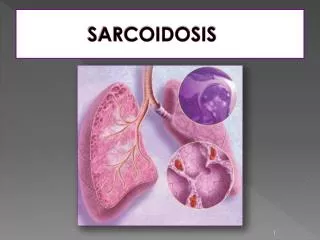
INTRODUCTION • Sarcoidosis is a systemic disorder of unknown cause with a wide variety of clinical and radiologic manifestations. • The diagnosis is usually made on the basis of these manifestations supported by histologic findings (noncaseating granulomes with proliferation of epithelioid cells) • Systemic manifestations (eg, Lofgren syndrome, Heerfordt syndrome) are commonly seen at clinical examination. Bilateral hilarlymphadenopathy is the most common radiologic finding. However, extra thoracic involvement can be an initial manifestation of symptomatic patients. • Familiarity with the clinical and radiologic features of sarcoidosis in various anatomic locations plays a crucial role in diagnosis and management.
OBJECTIVES • In this Study, we review sarcoidosis in terms of clinical and radiologic manifestations in a variety of organs. • We illustrate through some observations rare localization of sarcoidosis and discuss the contribution of imaging.
Material and methods • A series of nine patients collected over 4 years in MT Maamouri hospital, Nabeul, Tunisia. • The diagnosis of sarcoidosis is established on the basis of: * Clinical and radiologic findings * Histologic findings: Lung biopsy (transbronchial) Extrapulmonary sites (cervical lymph node and liver biopsy )
Results and discussion • Average age: 41 years • Sex ratio: 8 F/ 1 M • Clinical signs and symptoms are non specific and include: • cough • headache • polyuriapoldipsia syndrome • depression, dizziness • bone pain • skin lesions • abdominal pain • Biology shows: • Inflammatory syndrome in all patients. • Hypercalcaemia and elevated angiotensin converting enzyme are noted in 5 patients.
Results and discussion Various locations have been reported: • Mediastinal and/or lungs involvements (n=8) • Neurosarcoidosisinvolving: . Hypothalamic-pituitary axis (n=2) . Brain parenchymal localization (n=1) . Leptomeningeal involvement (n=2) • Hepaticsarcoidosis: . Homogeneous hepatomegaly (n=2) . Nodular form (n=2) • Splenic localization: . Homogenous splenomegaly (n=2) • Thyroid involvement (n=1) • PeriphericBone localization (n=2)
ThoracicInvolvement • Pulmonary involvement is reported to about 90% of our patients. There are five radiologic stages of intrathoracic changes: • Stage 0: Normal chest radiograph; • Stage 1: Lymphadenopathy only • Stage 2: Lymphadenopathy with parenchymal infiltration • Stage 3: Parenchymal disease only • Stage 4: Pulmonary fibrosis. One-half of patients present with stage 1 disease.
Chest radiograph of stage 1 sarcoidosis Patient has hilaradenopathyand no evidence of parenchymallunginvolvment. Chest radiograph of stage 3sarcoidosis Patient has diffuse lung disease but no significant adenopathy.
MediastinalLymphNodes • Intrathoraciclymphadenopathy is the most commonly encountered radiologic finding in sarcoidosis (80% of cases) and typically manifests as bilateral hilaradenopathy with right paratrachealadenopathy. • Contrast enhanced (CT) scan clearly depicts the bilateralhilaradenopathy.
LUNGS INVOLVEMENTS • Lung involvement in sarcoidosis is seen approximately 20% of patients and it has a strong predilection for the upperlung. • At histologic analysis, sarcoid granulomas in the lung are typically distributed along the lymphatic vessels, which run within the interstitial tissues of bronchovascular bundles and the subpleural and perilobular spaces .
High-resolution CT can accurately depict this characteristic distribution and typically demonstrates multiple small nodules in a perivascular distribution, along with irregular thickening of bronchovascular bundles and interlobularsepta. Pulmonary sarcoidosis in a 37-year-old man HR chest CT scan demonstrates: • * Small nodules with a Perivascular distribution • and along the interlobular pleura. • * Irregular thickening of bronchovascular bundles.
NEUROSARCOIDOSIS • Neurosarcoidosis has a strong predilection for Leptomeningesespecially observed the base of the brain. • Cranial nerve involvement, especially facial nerve, the optic nerve and chiasm are also frequently involved. • Neuroendocrine disorderssuch as diabetes insipidus that arise owing to involvement of the pituitary gland and hypothalamus are relatively common.
LEPTOMENINGEAL INVOLVEMENT Coronal and axial contrast-enhanced T1-weighted MR image show diffuselyenhancingleptomeningeallesions ( arrows)
HYPOTHALAMIC-PITUITARY AXIS coronal T1-weighted MR image reveal an enlarged hypothalamus
CRANIAL NERVE INVOLVEMENT On a contrast-enhanced T1-weighted MRimage • 1- The V Cranial Nerve enhancement • 2- The optic chiasm enhancement.
SPINAL CORD • T2-weighted MR images demonstrate an intramedullary hyperintense lesion • Contrast-enhanced T1-weighted MR image depicts an leptomeningealenhancing.
INTRA AXIAL BRAIN LESION contrast-enhanced T1-weighted MR image shows a an enhancing intraaxiallesion
PACHYMENINGEAL INVOLVEMENT Coronal / Sagital contrast-enhanced T1-weighted MR image show enhancing covering the Tentoriumcerebelli.
HEPATIC AND SPLENIC SARCOIDOSIS • Liverenlargementisfound on ultrasound or CT scan in up to 40% of cases . It’s the commonest CT finding, oftenaccompagnedwithsplenomegaly and (lessoften) abdominal lymphnodesenlargement. • Contrast-enhanced abdominal CT scan show homogenous organomegaly .
HEPATIC AND SPLENIC SARCOIDOSIS • Hepaticgranulomasare found on CT in only few cases, typicallyvisualized as multiple, discrete, lowattenuating, non enhancing (aftercontrast injection) nodules of variable size. • Simultaneous Splenicnodules favors a diagnosis of sarcoidosis. • Hepatic involvement in • A 49-year-old woman • with Pulmonary • sarcoidosis. • Contrast-enhanced • abdominal CT scan • shows multiple, • irregularly shaped • nodules of variable • size in the liver.
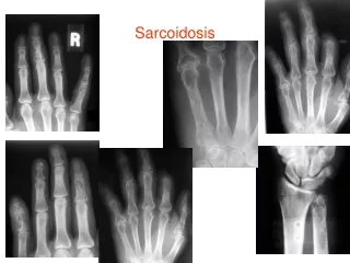
O S S E O U S S A R C O I D O S I S • Skeletal involvement is seen in approximately 10% of patients with sarcoidosis. • The phalanges in the hands and feet are most frequently affected. • These lesions are often multiple Radiologic features may include: - cystlike radiolucent areas - a lacelike honeycomb appearance - extensive bone erosion with pathologic fractures. • A subcutaneous soft-tissue mass, swelling or tenosynovitis may also be present. • The articular spaces are usually intact, unless extensive neuropathiclesionsDevelop.
O S S E O U S S A R C O I D O S I S • a radiograph of the right hand reveals a radiolucent lesion inthe proximal phalanges of the fourth and fifth digitis. • The lesion has a lacelike appearance and is accompanied by a soft-tissue swelling
O S S E O U S S A R C O I D O S I S • The distal phalanges of the right third and leftfourthtoespresentsimilartrabucular architecture and bonyremodeling changes. • On the distal phalanges of bothhalluces minute cortical defects are demonstrated. • The joints spacewidthis normal
THYROID INVOLVEMENT • Involvement of the thyroid gland occurs in 6% of patients with sarcoidosis. It is commonly associated with widespread disease in other organs. • Thyroid ultrasound in 59- years • woman shows diffuse enlargement • With isohypo-echoid • Heterogenousnoduls. • Thyroid biopsy Specimens after • loboisthmectomy demonstrates a • noncaseatinggranulomas with • epithelioid cells. • Laboratory data show that the • angiotensin-converting enzyme • (ACE) level is commonly elevated. • Salivary glands biopsy shows the • same sarcoidosicGranulomas.
SUMMARY • Sarcoidosis is a multisystem disease and diagnosis must be made on radio-clinic laboratory findings combined with histological evidence of non caseatinggranulomas. • Because the disease frequently involves multiple organs, familiarity with the clinical and radiologic features of sarcoidosis in various anatomic locations plays a crucial role in diagnosis and management. • Research is underway to improve imaging to guide the diagnosis and lead to an early and accurate diagnosis.
references • Koyama T, Ueda H, Togashi K, Umeoka S, KataokaM,Nagai S. Radiologic manifestations of sarcoidosis in various organs. Radiographics 2004; 24:87-104. • Roberts SD, Mirowski GW, Wilkes D, Kwo PY, Knox KS. Sarcoidosis. Part II: extrapulmonary and systemic manifestations. J Am AcadDermatol 2004; 51: 628-630. • Baughman R. Pulmonary sarcoidosis, MD University of Cincinnati Medical Center, USA. Clin Chest 2004; 25: 521–530. • Kragiannidis A, Karavalki M, KoulaouzidisA. Hepatic sarcoidosis. Annals of Hepatology 2006; 5: 251-256. • Mana J. Magnetic resonance imaging and nuclear imaging in sarcoidosis. CurrOpinPulm Med 2002; 8: 457–463. • Neville E, Carstairs LS, James DG. Sarcoidosis of bone. Q J Med 1977; 46:215–227.