Download
1 / 78
830 likes | 1.38k Views
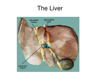
THE LIVER. Introduction/General Information A. Largest of viscera 1. ~ 2.5% body weight 2. Completely covered by Glisson’s Capsule 3. Incomplete covering by peritoneum. General Information, continued … 4. Measurements are ~ a. 21 – 23 cm transverse

E N D
Introduction/General Information • A. Largest of viscera • 1. ~ 2.5% body weight • 2. Completely covered by Glisson’s Capsule • 3. Incomplete covering by peritoneum
General Information, continued … 4. Measurements are ~ a. 21 – 23 cm transverse b. 15 – 18 cm superior to inferior c. 10 – 13 cm anterior to posterior
General Information, continued … Location: 1. right hypochondrium 2. epigastrium C. Mostly covered by ribs D. Contains numerous vascular structures
II. Detailed Anatomy • A. Four lobes • 1. Divisions based on blood supply, bile drainage • 2. Anatomical lobes divided by falciform ligament
Detailed Anatomy, con’t… 3. Functional Lobes a. right and left lobes separated by imaginary line b. from fossa for GB IVC
Right and Left Functional Lobes of the Liver Left Lobe Right Lobe
Detailed Anatomy, continued … • Functional Divisions • Right lobe with caudate process • Left lobe: • a. Caudate lobe • b. Quadrate lobe Caudate Process
Detailed Anatomy, continued … • C. Fissures: • 1. Right sagittal (main) • 2. Left sagittal (accessory) • 3. Portal • 4. Right oblique intersegmental • 5. Lateral intersegmental
Fissures of the Liver • Main lobar fissure • Boundary between R and L lobes • L.S. on U/S: seen as hyperechoic line from PV to neck of GB • Used to ID GB when it is packed with stones
Fissures of the Liver • Portal fissure • T.S. on U/S • Created by portal veins (triads) • R. main PV is // to anterior body wall
Segments of the Liver • Hepatic segments • I = caudate lobe • II & III = superior and inferior lateral segments, L. lobe • IV = medial segment, L. lobe • V & VI = caudal to transverse plane • VII & VIII = cephalad to transverse plane I
Functional divisions, continued … 4. Fossae (Superficial) a. IVC – posterior b. Portal Vein – inferior c. Gallbladder – inferior
Fossae, Inferior Surface of the Liver • Fossa for IVC • Fossa for Portal Vein • Fossa for Gall Bladder
Functional Divisions, continued … 5. Impressions (visceral surface): produced by abdominal viscera a. Gastric (fundus of stomach) b. Renal (right kidney) c. Adrenal (right adrenal gland) d. Duodenal (bulb of duodenum) e. Esophageal (esophagus) f. Right and left colic (flexures of the colon)
Visceral Impressions, continued … P • Esophageal • Renal • Gastric • Adrenal • Duodenal • Right colic • Left colic L R A
Detailed Anatomy, continued … D. Ligaments 1. Falciform (most superficial anteriorly) a. Divides left lobe in two sections 1. anatomical left lobe 2. caudate & quadrate lobes b. Two layers of peritoneum c. Extends to umbilicus
Ligaments, continued … 2. Ligamentum teres hepatis (fetal source??) 3. Ligamentum venosum (fetal source??) 4. Right/Left Coronary Ligaments
Hepatic Ligaments • Falciform ligament • L. coronary ligament • L. triangular ligament • Ligamentum teres hepatis • Ligamentum venosum
Ligaments, continued … • 5. Hepatophrenic & Hepatorenal ligaments: • a. Subdivisions of right coronary ligament • b. hepatophrenic (superior) & hepatorenal (inferior) • c. Surround BARE AREA
Hepatic Ligaments, con’t… • Hepatophrenic ligament • Hepatorenal ligament Bare Area
Detailed Anatomy, continued … E. Lesser Omentum 1. Sleeve-like structure 2. Connects lesser curvature of stomach & bulb of duodenum to inferior surface of liver 3. AKA: Gastrohepatic or Hepatoduodenal ligament
Lesser Omentum, continued … 3. Attachment surrounds Porta Hepatis 4. Continues on each side of ligamentum venosum 5. Extends to caudate & left lobes on posterior surface of liver
Lesser Omentum • Lesser Omentum: (R) Anterior view, (L) Inferior view
Detailed Anatomy, con’t… • E. Subphrenic Spaces • clinically important • common sites for abscesses • Between liver and diaphragm
Subphrenic Spaces, con’t… • 4. Right superior posterior subphrenic space • a. Boundaries: • - superior: right coronary ligament • - anterior: liver • - posterior: parietal peritoneum covering diaphragm • b. Extends inferiorly to….
Subphrenic Spaces, continued … 5. Right posterior inferior subphrenic space a. Boundaries - above: inferior surface of liver - below: transverse colon & mesocolon b. Extends over right adrenal & kidney
Subphrenic Spaces, continued … c. AKA: Hepatorenal Pouch/Recess, Morrison’s Pouch d. Patient lying supine: 1. Lowest part of peritoneal cavity is behind liver 2. Fluid, pus, etc. collects here 3. Can cause abscess formation
Subphrenic spaces, continued … • 6. Right superior anterior subphrenic space • a. Boundaries: • - right side of falciform ligament • - upper layer of right coronary ligament • - underside of diaphragm • - superior surface of liver • b. Found when patient lying prone
Subphrenic spaces, continued … 7. Most sources of peritoneal contamination are on the right 8. Right posterior & right inferior spaces are most significant sites 9. Infection may spread via diaphragmatic lymphatics
Detailed Anatomy, continued … • F. Porta Hepatis: contains the following structures • 1. Hepatic arteries • a. Usually two, sometimes one • b. Originate from common/proper hepatic artery • c. Course is variable
Porta Hepatis, continued … • 2. Portal vein (supplies 1100 ml/blood per minute) • a. Largest structure in porta • b. Formed by confluence of mesenteric veins & splenic vein
Porta Hepatis Hepatic Artery Portal Vein Common Bile Duct
Porta Hepatis, continued … • 3. Hepatic & cystic ducts • 4. Nerves: • a. Vagus X (parasympathetic) • b. Fibers from celiac ganglion
Porta Hepatis, continued … 4. Lymph nodes: a. most hepatic lymph vessels end in nodes around porta hepatis • b. From here, drain into celiac nodes
Porta Hepatis, continued … • c. Some vessels pass through falciform ligament • 1. through diaphragm • 2. into mediastinal nodes • d. enlarged nodes may compress portal vein or hepatic duct
Detailed Anatomy, continued … G. Vascular Structures in Liver 1. Largest vessels are portal vein and IVC a. Portal Vein: 1. appears on T.S. as tubular, echolucent structure 2. courses horizontally from porta hepatis
Detailed Anatomy, continued … 3. walls echogenic due to structures in portal triad b. Left Portal Vein: 1. has more variable course 2. May be difficult to trace on transverse scans
Hepatic Vessels • IVC • Right Portal Vein • Left Portal Vein • Main Portal Vein
Vascular Structures, continued … c. Right Portal Vein: 1. anatomical landmark 2. extends into right lobe 3. branches after porta hepatis 4. L.S. shows “dumbbell” or circular structure with echogenic “collar”
Vascular Structures, continued … d. IVC: 1. To right of aorta 2. Appears to pass through liver 3. Diameter enlarges after renal veins join (~L-1)
Vascular structures, continued … 2. Hepatic Veins: a. Tubular structures b. Enlarge cephalad c. In superior half of liver d. Angles of hepatic vein branches oriented toward IVC e. Walls not echogenic
Hepatic Veins R. Hepatic Vein Middle Hepatic Vein L. Hepatic Vein
Detailed Anatomy, continued … • Bile Ducts • 1. No “normal” anatomy • 2. If dilated: • a. Echogenic collar • b. Lobulated shape • c. Highly branched over short distances • d. Converge toward porta hepatis