Download
1 / 66
700 likes | 1.17k Views
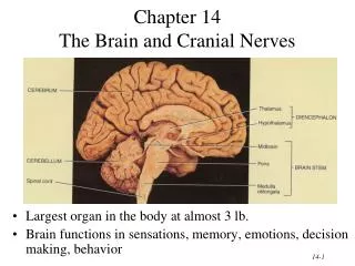
The Brain and Cranial Nerves. Largest organ in the body at almost 3 lb. Brain functions in sensations, memory, emotions, decision making, behavior. The Cerebrum. Figure 14–12a The Brain in Lateral View. Principal Parts of the Brain. Cerebrum Diencephalon thalamus & hypothalamus Cerebellum

E N D
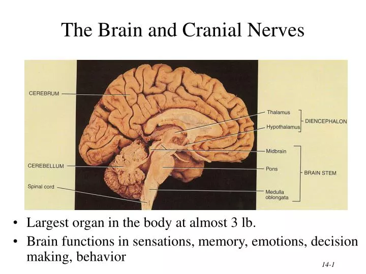
The Brain and Cranial Nerves • Largest organ in the body at almost 3 lb. • Brain functions in sensations, memory, emotions, decision making, behavior
The Cerebrum Figure 14–12a The Brain in Lateral View.
Principal Parts of the Brain • Cerebrum • Diencephalon • thalamus & hypothalamus • Cerebellum • Brainstem • medulla, pons & midbrain
Protective Coverings of the Brain • Bone, meninges & fluid • Meninges same as around the spinal cord • dura mater • arachnoid mater • pia mater
Brain Protection and Support Figure 21–23 Arteries of the Brain.
Blood Supply to Brain • Arterial blood supply is branches from circle of Willis on base of brain • Vessels on surface of brain----penetrate tissue • Uses 20% of our bodies oxygen & glucose needs • blood flow to an area increases with activity in that area • deprivation of O2 for 4 min does permanent injury • at that time, lysosome release enzymes • Blood-brain barrier (BBB) • protects cells from some toxins and pathogens • proteins & antibiotics can not pass but alcohol & anesthetics do • tight junctions seal together epithelial cells, continuous basement membrane, astrocyte processes covering capillaries
Cerebrospinal Fluid (CSF) • 80-150 ml (3-5oz) • Clear liquid containing glucose, proteins, & ions • Functions • mechanical protection • floats brain & softens impact with bony walls • chemical protection • optimal ionic concentrations for action potentials • circulation • nutrients and waste products to and from bloodstream
Origin of CSF • Choroid plexus = capillaries covered by ependymal cells • 2 lateral ventricles, one within each cerebral hemisphere • roof of 3rd ventricle • fourth ventricle
Drainage of CSF from Ventricles • One median aperture & two lateral apertures allow CSF to exit from the interior of the brain
Reabsorption of CSF • Reabsorbed through arachnoid villi • grapelike clusters of arachnoid penetrate dural venous sinus • 20 ml/hour reabsorption rate = same as production rate
Hydrocephalus • Blockage of drainage of CSF (tumor, inflammation, developmental malformation, meningitis, hemorrhage or injury • Continued production cause an increase in pressure --- hydrocephalus • In newborn or fetus, the fontanels allow this internal pressure to cause expansion of the skull and damage to the brain tissue • Neurosurgeon implants a drain shunting the CSF to the veins of the neck or the abdomen
Medulla Oblongata • Continuation of spinal cord • Ascending sensory tracts • Descending motor tracts • Nuclei of 5 cranial nerves • Cardiovascular center • force & rate of heart beat • diameter of blood vessels • Respiratory center • medullary rhythmicity area sets basic rhythm of breathing • Information in & out of cerebellum • Reflex centers for coughing, sneezing, swallowing etc
Ventral Surface of Medulla Oblongata • Ventral surface bulge • pyramids • large motor tract • decussation of most fibers • left cortex controls right muscles • Olive = olivary nucleus • neurons send input to cerebellum • proprioceptive signals • gives precision to movements
Dorsal Surface of Medulla Oblongata • Nucleus gracilis & nucleus cuneatus = sensory neurons • relay information to thalamus on opposite side of brain • 5 cranial nerves arise from medulla -- 8 thru 12
XII = Hypoglossal Nerve • Controls muscles of tongue during speech and swallowing • Injury deviates tongue to injured side when protruded • Mixed, primarily motor
XI = Spinal Accessory Nerve • Cranial portion • arises medulla • skeletal mm of throat & soft palate • Spinal portion • arises cervical spinal cord • sternocleidomastoid and trapezius mm.
X = Vagus Nerve • Receives sensations from viscera • Controls cardiac muscle and smooth muscle of the viscera • Controls secretion of digestive fluids • 90% Parasympathetics
Cranial Nerves Figure 14–26 The Vagus Nerve.
IX = Glossopharyngeal Nerve • Stylopharyngeus m. (lifts throat during swallowing) • Secretions of parotid gland • Somatic sensations & taste on posterior 1/3 of tongue
VIII = Vestibulocochlear Nerve • Cochlear branch begins in medulla • receptors in cochlea • hearing • if damaged deafness or tinnitus (ringing) is produced • Vestibular branch begins in pons • receptors in vestibular apparatus • sense of balance • vertigo (feeling of rotation) • ataxia (lack of coordination)
Cranial Nerves Figure 14–24 The Vestibulocochlear Nerve.
Pons • One inch long • White fiber tracts ascend and descend • Pneumotaxic & apneustic areas help control breathing • Middle cerebellar peduncles carry sensory info to the cerebellum • Cranial nerves 5 thru 7
VII = Facial Nerve • Motor portion • facial muscles • salivary & nasal and oral mucous glands & tears • Sensory portion • taste buds on anterior 2/3’s of tongue
VI = Abducens Nerve • Lateral rectus eye muscle
V = Trigeminal Nerve • Motor portion • muscles of mastication • Sensory portion • touch, pain, & temperature receptors of the face • ophthalmic branch • maxillary branch • mandibular branch
Cranial Nerves Figure 14–22 The Trigeminal Nerve.
Midbrain • One inch in length • Extends from pons to diencephalon • Cerebral aqueduct connects 3rd ventricle above to 4th ventricle below
Midbrain in Section • Cerebral peduncles---clusters of motor & sensory fibers • Substantia nigra---helps controls subconscious muscle activity • Red nucleus-- rich blood supply & iron-containing pigment • cortex & cerebellum coordinate muscular movements by sending information here from the cortex and cerebellum
Dorsal Surface of Midbrain • Corpora quadrigemina = superior & inferior colliculi • coordinate eye movements with visual stimuli • coordinate head movements with auditory stimuli
IV = Trochlear Nerve • Superior oblique eye muscle
III = Oculomotor Nerve • Levator palpebrae raises eyelid (ptosis) • 4 extrinsic eye muscles • 2 intrinsic eye muscles • accomodation for near vision (changing shape of lens during reading) • constriction of pupil
Cranial Nerves Figure 14–21 Cranial Nerves Controlling the Extra-Ocular Muscles.
Lecture 2 Reticular Formation • Scattered nuclei in medulla, pons & midbrain • Reticular activating system • alerts cerebral cortex to sensory signals (sound of alarm, flash light, smoke or intruder) to awaken from sleep • maintains consciousness & helps keep you awake with stimuli from ears, eyes, skin and muscles • Motor function is involvement with maintaining muscle tone
Cerebellum • 2 cerebellar hemispheres and vermis (central area) • Function • correct voluntary muscle contraction and posture based on sensory data from body about actual movements • sense of equilibrium
Cerebellum • Transverse fissure between cerebellum & cerebrum • Cerebellar cortex (folia) & central nuclei are grey matter • Arbor vitae = tree of life = white matter
Thalamus • 1 inch long mass of gray mater in each half of brain (connected across the 3rd ventricle by intermediate mass) • Relay station for sensory information on way to cortex • Crude perception of some sensations
Hypothalamus • Major regulator of homeostasis • receives somatic and visceral input, taste, smell & hearing information; monitors osmotic pressure, temperature of blood
Functions of Hypothalamus • Controls and integrates activities of the ANS which regulates smooth, cardiac muscle and glands • Synthesizes regulatory hormones that control the anterior pituitary • Contains cell bodies of axons that end in posterior pituitary where they secrete hormones • Regulates rage, aggression, pain, pleasure & arousal • Feeding, thirst & satiety centers • Controls body temperature • Regulates daily patterns of sleep
Epithalamus • Pineal gland • endocrine gland the size of small pea • secretes melatonin during darkness • promotes sleepiness & sets biological clock • Habenular nuclei • emotional responses to odors
Cerebrum (Cerebral Hemispheres) • Cerebral cortex is gray matteroverlying white matter • 2-4 mm thick containing billionsof cells • grew so quickly formed folds(gyri) and grooves (sulci or fissures) • Longitudinal fissure separates left & right cerebral hemispheres • Corpus callosum is band of white matter connecting left and right cerebral hemispheres • Each hemisphere is subdivided into 4 lobes
Longitudinal fissure (green) Frontal lobe Central sulcus (yellow) precentral & postcentral gyrus Parietal lobe Parieto-occipital sulcus Occipital lobe Lateral sulcus (blue) Temporal lobe Insula Lobes and Fissures
The Cerebrum Figure 14–12b The Brain in Lateral View.
The Cerebrum Figure 14–13b Fibers of the White Matter of the Cerebrum.
Basal Ganglia • Connections to red nucleus, substantia nigra & subthalamus • Input & output with cerebral cortex, thalamus & hypothalamus • Control large automatic movements of skeletal muscles
Limbic System • Parahippocampal & cingulate gyri & hippocampus • Emotional brain--intense pleasure & intense pain • Strong emotions increase efficiency of memory
Brain Injuries • Causes of damage • displacement or distortion of tissue at impact • increased intracranial pressure • infections • free radical damage after ischemia • Concussion---temporary loss of consciousness • headache, drowsiness, confusion, lack of concentration • Contusion--bruising of brain (less than 5 min unconsciousness but blood in CSF) • Laceration--tearing of brain (fracture or bullet) • increased intracranial pressure from hematoma
Sensory Areas of Cerebral Cortex Receive sensory information from the thalamus Primary somatosensory area = postcentral gyrus = 1,2,3 Primary visual area = 17 Primary auditory area = 41 & 42 Primary gustatory area = 43
The Cerebrum Figure 14–15a Motor and Sensory Regions of the Cerebral Cortex.
Motor Areas of Cerebral Cortex • Voluntary motor initiation • Primary motor area = 4 = precentral gyrus • controls voluntary contractions of skeletal muscles on other side • Motor speech area = 44 = Broca’s area • production of speech -- control of tongue & airway