Download
1 / 67
740 likes | 1.19k Views
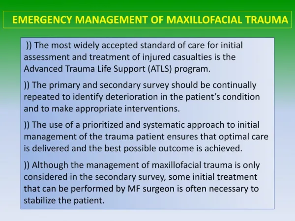
Maxillofacial Trauma. Joe Lex, MD, FACEP, FAAEM Temple University School of Medicine Philadelphia, PA USA Joseph.Lex@TUHS.Temple.edu. Lecture Outline. Emergency management Facial exam Fractures Major Minor Soft tissue injuries Unusual injuries. FACE. Causes of Mortality. Acute

E N D
Maxillofacial Trauma Joe Lex, MD, FACEP, FAAEM Temple University School of Medicine Philadelphia, PA USA Joseph.Lex@TUHS.Temple.edu
Lecture Outline • Emergency management • Facial exam • Fractures • Major • Minor • Soft tissue injuries • Unusual injuries FACE
Causes of Mortality • Acute • Airway compromise • Exsanguination • Associated intracranial or cervical-spine injury • Delayed • Meningitis • Oropharyngeal infections FACE
Epidemiology • Estimated 3,000,000 facial trauma cases per year in USA • Estimated 40 to 50% of motor vehicle victims have facial injury • No uniform reporting or registry of cases FACE
Functions of Face • Respiratory upper airway • Visual • Olfactory • Mastication • Cosmetic • Communication • Individual recognition FACE
Management Sequence • Airway control / immobilize cervical spine • Bleeding control • Complete the primary survey • Secondary survey • Consider NG or OG tube placement FACE
Management Sequence • Plain radiographs if fractures suspected • CT if suspect complex fractures FACE
Management Sequence • Repair soft tissue immediately if no other injuries • Delay soft tissue repair until patient in OR if surgery for other injuries necessary FACE
Initial Management Step 1: Airway control • Oxygen for all patients • May need to keep patient sitting or prone • Stabilize C-spine early • Large bore (Yankauer) suction available FACE
Initial Management Step 1: Airway control • Orotracheal intubation preferred over nasotracheal if possible midfacial fracture and invasive airway needed • Combitube®, retrograde wire, or cricothyroidostomy if unable to orotracheally intubate FACE
Initial Management Step 2 : Bleeding control • Can be major threat to life • Use universal precautions • Direct pressure dressings initially • Contraindicated: blind vessel clamping FACE
Initial Management Step 2 : Bleeding control • Rapid nasal packing may be necessary • Be sure blood is not just running down posterior pharynx FACE
Initial Management Step 2 : Bleeding control • Rarely: emergent cutdown and ligation of external carotid artery needed to prevent exsanguination • Note: Although shock in facial trauma patient is usually due to other injuries, it is possible to bleed to death from a facial injury FACE
Airway Compromise • Blood in airway • “Debris” in airway • Vomitus, avulsed tissue, teeth or dentures, foreign bodies • Pharyngeal or retropharyngeal tissue swelling • Posterior tongue displacement from mandible fractures FACE
Secondary Survey Scalp • Check for lacerations, hematomas, stepoffs, tenderness • Bleeding maybe brisk until sutured • Can use stapler for rapid closure FACE
Secondary Survey Ears • Examine pinnae, canal walls, tympanic membranes • Suction gently under direct vision if blood in canal • Put drop of canal fluid on filter paper for “ring sign” CSF leak • Assess hearing FACE
Secondary Survey Eyes • Pupils, anterior chamber, fundi, extraocular movements • Conjunctivae for foreign bodies • Palpate orbital rims • No globe palpation if suspect penetration FACE
Secondary Survey Eyes • Lid injury can leave cornea exposed • Use artificial tears or cellulose gel FACE
Secondary Survey Overall facial appearance • Assess for symmetry, deformity, discoloration, nasal alignment • Palpate forehead & malar areas FACE
Secondary Survey Nose • Check septum for hematoma & position • Check airflow in both nares • Palpate nasal bridge for crepitus • Check fluid on filter paper for “ring sign” (for CSF leak) FACE
Secondary Survey Mouth • Check occlusion • Reflect upper & lower lips • Check Stenson's duct for blood • Palpate along mandibular and maxillary teeth (be careful !) FACE
Secondary Survey Mouth • Palpate along exterior of mandible • Pull forward on maxillary teeth FACE
Secondary Survey Neurologic • Skin fold symmetry at rest • Motor: each division of CN-VII • Sensation: 3 divisions of CN-V • Sensation on tongue • Gag reflex FACE
Major Lefort I, II, III Mandibular Minor Nasal Sinus wall Zygomatic Orbital floor Antral wall Alveolar ridge Fracture Classification FACE
Forces Required • Nasal fracture 30 g • Zygoma fractures 50 g • Mandibular (angle) fractures 70 g • Frontal region fractures 80 g • Maxillary (midline) fractures 100 g • Mandibular (midline) fractures 100 g • Supraorbital rim fractures 200 g FACE
Lefort Fractures • Lefort fractures can coexist with additional facial fractures • Patient may have different Lefort type fracture on each side of the face FACE
Differentiating Leforts Pull forward on maxillary teeth • Lefort I: maxilla only moves • Lefort II: maxilla & base of nose move: • Lefort III: whole face moves: FACE
Lefort I: Nasomaxillary • Horizontal fracture extending through maxilla between maxillary sinus floor & orbital floor • Crepitus over maxilla • Ecchymosis in buccal vestibule • Epistaxis: can be bilateral • Malocclusion • Maxilla mobility FACE
Lefort I: Nasomaxillary • Closed reduction • Intermaxillary fixation: secures maxilla to mandible • May need wiring or plating of maxillary wall and / or zygomatic arch • Antibiotics: anti-staphylococcal FACE
Lefort II: Pyramidal • Subzygomatic midfacial fracture with a pyramid-shaped fragment separated from cranium and lateral aspects of face FACE
Lefort II: Pyramidal Signs & symptoms • Midface crepitus • Face lengthening • Malocclusion • Bilateral epistaxis • Infraorbital paresthesia • Ecchymoses: buccal vestibule, periorbital, subconjunctival FACE
Lefort II: Pyramidal • Hemorrhage or airway obstruction may require emergent surgery • Treatment can often be delayed till edema decreased FACE
Lefort II: Pyramidal Usually require • Intermaxillary fixation • Interosseous wiring or plating of infraorbital rims, nasal-frontal area, & lateral maxillary walls • May need additional suspension wires • Antibiotics FACE
Lefort III • Craniofacial dissociation • Bilateral suprazygomatic fracture resulting in a floating fragment of mid-facial bones, which are totally separated from the cranial base FACE
Lefort III Signs and Symptoms • Face lengthening: “caved-in” or “donkey face” • Malocclusion: “open bite” • Lateral orbital rim defect • Ecchymoses: periorbital, subconjunctival FACE
Lefort III Signs and Symptoms • Bilateral epistaxis • Infraorbital paresthesia • Often medial canthal deformity • Often unequal pupil height FACE
Lefort III • Usually associated with major soft tissue injury requiring emergent surgery for bleeding control • Surgery can be delayed till edema resolves • Intermaxillary fixation FACE
Lefort III • Transosseous wiring or plating • Frontozygomatic suture • Nasofrontal suture • May need extracranial fixation if concurrent mandibular fracture • Antibiotics FACE
Forces Required • Nasal fracture 30 g • Zygoma fractures 50 g • Mandibular (angle) fractures 70 g • Frontal region fractures 80 g • Maxillary (midline) fractures 100 g • Mandibular (midline) fractures 100 g • Supraorbital rim fractures 200 g FACE
Mandible Fractures • Airway obstruction from loss of attachment at base of tongue • >50 % are multiple • Condylar fractures associated with ear canal lacerations & high cervical fractures • High infection potential if any violation of oral mucosa FACE
Mandible Fractures Signs and symptoms • Malocclusion • Decreased jaw range of motion • Trismus • Chin numbness • Ecchymosis in floor of mouth • Palpable step deformity FACE
Mandible Fractures • Tongue blade test: have patient bite down while you twist. If no fracture, you will be able to break the blade. FACE
Mandible Fractures Treatment • Prompt fixation: intermaxillary fixation (arch bars), +/- body wiring or plating FACE
TMJ Dislocation • Can occur from direct blow to mandible • Can occur “spontaneously” from yawning or laughing • Mandible dislocates forward & superiorly • Concurrent masseter & pterygoid spasm FACE
TMJ Dislocation Symptoms • Patient presents with mouth open, cannot close mouth or talk well • Can be misdiagnosed as psychiatric or dystonic reaction FACE
TMJ Dislocation Treatment • Manual reduction: place wrapped thumbs on molars & push downward, then backward • Be careful not to get bitten • Usually does not require procedural sedation or muscle relaxants FACE
Forces Required • Nasal fracture 30 g • Zygoma fractures 50 g • Mandibular (angle) fractures 70 g • Frontal region fractures 80 g • Maxillary (midline) fractures 100 g • Mandibular (midline) fractures 100 g • Supraorbital rim fractures 200 g FACE
Nasal Bone Fractures • Often diagnosed clinically: x-ray not needed • Emergent reduction not necessary except to control epistaxis • Usually do not need antibiotics • Early reduction under local anesthesia useful if nares obstructed FACE
Nasal Bone Fractures • Nasal septal hematoma: incise & drain, anterior pack, antibiotics, follow-up at 24 hours • Follow-up timing for recheck or reduction: • Children: 3 to 5 days • Adults: 7 days FACE
Forces Required • Nasal fracture 30 g • Zygoma fractures 50 g • Mandibular (angle) fractures 70 g • Frontal region fractures 80 g • Maxillary (midline) fractures 100 g • Mandibular (midline) fractures 100 g • Supraorbital rim fractures 200 g FACE