Download
1 / 26
350 likes | 707 Views
بسم الله الرحمن الرحيم. Hormonal Changes in Pregnancy. Dr.Mohammed Sharique Ahmed Quadri Assistant professor physiology Al M aarefa College. Fertilization. Normal site of fertilization is ampulla of oviduct Must occur within 24 hours after ovulation Viability of ovum – 24 hrs .

E N D
بسم الله الرحمن الرحيم Hormonal Changes in Pregnancy Dr.Mohammed Sharique Ahmed Quadri Assistant professor physiology Al Maarefa College
Fertilization • Normal site of fertilization is ampulla of oviduct • Must occur within 24 hours after ovulation • Viability of ovum – 24 hrs. • Sperm usually survive about 48 hours but can survive up to 5 days in female reproductive tract • Sperm deposited in vagina travel through cervical canal, uterus, and to upper third of oviduct • Female reproductive tract aids in sperm migration • Contractions of myometrium • Upward contractions of oviduct smooth muscle
Fertilization • First sperm to reach ovum • Sperm binds with specific site on zona pellucida on ovum • The sperm undergo the “Acrosomalreaction” and secretes powerful enzymes that helps it to pierce through the corona radiata and zona pellucida of the ovum. • Fuses with plasma membrane of ovum • This fusion Triggers chemical change in ovum’s surrounding membrane that makes outer layer impermeable to entry of any more sperm • Head of fused sperm gradually pulled into ovum’s cytoplasm • Within hour, sperm and egg nuclei fuse • Fertilized ovum now called a zygote
Fertilization and Implantation • Fertilized ovum( zygote ) divides mitotically • Within week zygote grows and differentiates into blastocyst capable of implantation • The blastocyst implants in the posterior wall of uterus on 6-7th day after fertilization. • The blastocyst has inner cell mass that becomes embryo and outer cellular layer, Trophoblast (fetal portion of placenta). • At site of implantation Endometrium transforms into Decidual layer.(maternal portion of placenta).
Early Stages of Development from Fertilization to Implantation
Fertilization and Implantation • Blastocyst implants in endometrial lining by means of enzymes released by trophoblasts
Formation of placenta • After implantation Placenta develops • Placenta forms link between the foetus and mother. • Trophoblast forms fetal part • Decidual layer of the endometrium forms the maternal part • Trophoblast differentiates into Outer Syncytiotrophoblast and Inner Cytotrophoblast
functions of Placenta • Organ of exchange between maternal and fetal blood • Acts as transient, complex endocrine organ that secretes essential pregnancy hormones • Human chorionic gonadotropin( HCG) • Estrogen • Progesterone • Human Chorionic Somatotrophin:
Human chorionic gonadotropin(HCG) • Peptide hormone secreted by Syncytiotrophoblast • Similar to LH • Appears in blood within 1 week and urine within 2 weeks of conception. • Reaches peak at 8 weeks then declines Actions : • Similar to LH. Maintains Corpus Luteum of pregnancy to secrete Progesterone. At 8-10 weeks Placenta is fully developed and takes over the secretion of Progesterone. • In male fetus-stimulate secretion of testosterone • Presence in urine forms the basis of pregnancy test.
ESTROGEN • 1st trimester – secreted from Corpus Luteum • After that Placenta secretes Estriol. • Why developing placenta doesn’t produce estrogen? • As Placenta lacks certain enzymes needed for synthesis of estriol • formation of Estriol requires the participation of Fetal Adrenal cortex together with Placenta. Actions During Pregnancy:- • Stimulates growth of myometrium--increasing uterine strength for parturition • Promotes development of ducts in mammary gland-prepare for lactation .
secretion of estrogen & progesterone from placenta The Feto-Placental Unit
PROGESTERONE • 1st Trimester – secreted from Corpus Luteum: after that from Placenta. Actions During Pregnancy – • Inhibits uterine contractions“Hormone of pregnancy”. • Stimulates alveolar development in Mammary gland- prepares for lactation . • Promotes formation of mucus plug in cervical canal – prevention of uterine contamination.
Human Placental Lactogen(HPL) /Human Chorionic Somatotrophin • Protein hormone secreted by syncytiotrophoblast • Similar in structure to GH and Prolactin Actions – • Mobilizes nutrients from mother to fetus. • (Reduces maternal use of glucose and promotes the breakdown of stored fat(similar to growth hormone) so that greater quantities of glucose and free fatty acids may be shunted to the fetus) • Helps prepare the mammary glands for lactation (similar to prolactin)
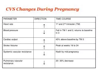
Physical changes within mother to meet demands of pregnancy • Period of gestation -About 38 weeks from conception. • Uterine enlargement • Breasts enlarge and develop ability to produce milk • Volume of blood increases 30% • Weight gain(Increased upto 12 Kgs) • Increased Heart Rate & Cardiac Output – 30-40% • Respiratory activity increases by about 20% • Urinary output increases • Kidneys excrete additional wastes from fetus • Nutritional requirements increase
Parturition • Labor, delivery, birth • Requires • Dilation of cervical canal to accommodate passage of fetus from uterus through vagina and to the outside • Contraction of uterine myometrium that are sufficiently strong to expel fetus • Exact factors triggering increase in uterine contractility and initiating parturition not fully established
Factors that triggers the onset of labor • Role of high Estrogen levels: Rising estrogen level with gestation • Makes the uterus more excitable by • Increase the number of gap junctions between myometrial cells • Increases oxytocin receptors in myometrium • contributes to cervical softening by • Increase local prostaglandin production
Factors that triggers the onset of labor • Role of corticotropin releasing hormone(CRH): • secreted by the fetal portion of the placenta into both the maternal and fetal circulations • when a critical level of placental CRH is reached, parturition is triggered.
Factors that triggers the onset of labor • Role of Oxytocin: Produce by hypothalamus, & released by posterior pituitary on neural stimulation • A powerful uterine muscle stimulant • Uterine responsiveness to oxytocin is 100 times greater at term than in nonpregnant women (because of the increase in gap junctions and increased concentration of myometrial oxytocin receptors) • labor is initiated when the myometrial responsiveness to oxytocin reaches a critical threshold that permits the onset of strong, coordinated contractions in response to ordinary levels of circulating oxytocin.
Parturition • Once contractions begin at labor onset, positive-feedback cycle progressively increases force of contraction • Pressure of fetus against cervix reflexly increases oxytocin secretion. • Which further pushes the fetus against the cervix • This cycle is reinforced as oxytocin stimulates prostaglandin secretion by decidua.
Stages of labor • Cervical dilation ( first stage) • Longest stage • Lasts from several hours to as long as 24 hours in a first pregnancy • Delivery of baby(second stage) • Begins when cervical dilation is complete • Stretching of vagina causes reflex contraction of abdominal muscles • Usually lasts 30 to 90 minutes • Delivery of placenta( third stage) • Second series of uterine contractions separates placenta from myometrium • Shortest stage – usually completed within 15 to 30 minutes after baby is born • After delivery, uterus shrinks to pregestational size (involution) in 4-6 week
References • Human physiology by Lauralee Sherwood, seventh edition • Text book physiology by Guyton &Hall,11th edition • Text book of physiology by Linda .s contanzo,third edition