Download
1 / 29
290 likes | 399 Views
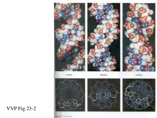
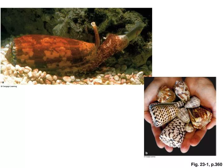
Fig. 23-1, p.360. Table 23-1, p.361. dorsal. posterior. ventral. anterior. Fig. 23-2, p.361. Fig. 23-3, p.363. placozoans. sponges. cnidarians. flatworms. rotifers. mollusks. annelids. roundworms. arthropods. echinoderms. chordates. deuterostomes, anus forms first in embryos.

E N D
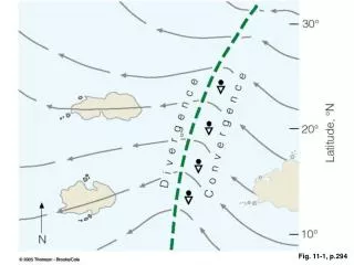
dorsal posterior ventral anterior Fig. 23-2, p.361
placozoans sponges cnidarians flatworms rotifers mollusks annelids roundworms arthropods echinoderms chordates deuterostomes, anus forms first in embryos protostomes, mouth forms first in embryos radial ancestry, two germ layers bilateral ancestry, three germ layers no true tissues true tissues multicelled body choanoflagellates fungi Fig. 23-6, p.364
water out glassy structural elements ameboid cell pore central cavity semifluid matrix flattened surface cells collar cell water in water in flagellum collar of microvilli nucleus Fig. 23-8, p.365
barbs on discharged thread exposed capsule's trigger (modified cilium) lid barbed thread in capsule nematocyst (capsule at free surface of epidermal cell) Fig. 23-10, p.366
reproductive polyp male medusa female medusa sperm ovum zygote feeding polyp one branch of a colony growth of a polyp ciliated bilateral larva Fig. 23-12, p.367
rudimentary brain (pair of large ganglia in head) ovary branching gut testis oviduct pharynx; protrudes onto food, then retracts into the body between feedings pair of nerve cords that have lateral branchings genital pore Fig. 23-13, p.368
proglottids scolex A human, the definitive host, eats infected, undercooked beef, which is mainly skeletal muscle. Larvae, each with inverted scolex of future tapeworm, become encysted in intermediate host tissues (e.g., skeletal muscle). scolex attached to wall of intestine one proglottid Inside each fertilized egg, an embryonic, larval form develops. Cattle may ingest embryonated eggs or ripe proglottids, and so become intermediate hosts. Each sexually mature proglottid has female and male organs. Ripe proglottids containing fertilized eggs leave the host in feces, which may contaminate water and vegetation. Fig. 23-15, p.369
esophagus stomach brain kidney arm jaw cuttlebone radula mantle accessory heart anus ink sac heart reproductive organ tentacle gill siphon Fig. 23-21, p.373
pharynx intestine eggs in uterus gonad false coelom (unlined body cavity) anus muscular body wall Fig. 23-22, p.374
chelicerae Fig. 23-25, p.376