Download
1 / 17
250 likes | 2.31k Views
Diabetic Macular Edema: What is Focal and What is Diffuse? . Sponsored by the National Eye Institute, National Institutes of Health, U.S. Department of Health and Human Services. Intravitreal Triamcinolone vs. Laser for DME. Participants Center involved DME (CST ≥250 um) Primary Outcome*

E N D
Diabetic Macular Edema: What is Focal and What is Diffuse? Sponsored by the National Eye Institute, National Institutes of Health, U.S. Department of Health and Human Services.
Intravitreal Triamcinolone vs. Laser for DME Participants Center involved DME (CST ≥250 um) Primary Outcome* Laser treated eyes had greater VA outcomes Secondary Outcome CST paralleled VA results *Ophthalmology 2008 Sep;115(9):1447-1459.e10
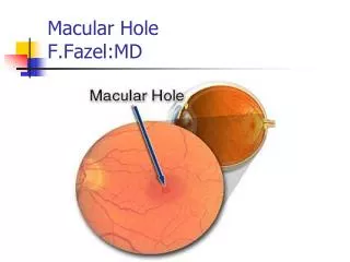
Does the treatment interventionhave the same response across subgroups? Secondary analysis subgroup participants into categories that may have different natural course Focal vs. Diffuse DME Focal DME Diffuse DME 3
Why Bother with Focal and Diffuse Diabetic Macular Edema? Background: “Focal edema responds BEST to laser” “Diffuse edema has WORSE natural course” “Diffuse edema responds BEST to steroids” Do we know what Focal or Diffuse DME is???
DRCR.net Perspective on Focal vs. Diffuse DME: Am J Ophthalmol 2008;146:649-55 Frequently used terms Attempt to differentiate 2 distinct types of DME, but………… • Do these terms have consistent definitions? • NOT REALLY!
Multiple Sources of Confusion Many articles discuss Focal/Diffuse without providinganydefinition Many say Diffuse = diffuse leakage; Focal = focal leakage Focal laser = direct treatment of MAs Published definitions use 4 exam methods, singly or in combination: Biomicroscopy, photos, FA, OCT
Published Definitions Vary Widely in Specific Criteria In general, definitions based on fundus photos mirror characteristics of definitions based on clinical exam alone
Published Definitions Vary Widely in Specific Criteria: Exam/Photo Area component ? ≥ 2 DA retinal thickening = diffuse (inconsistent cut points) Do we reliably quantitate area? Location component Foveal = diffuse Require area + location or only 1?? Lipid Lipid = focal Quantity (lots), Configuration (ring), or both
ETDRS classified proportion of FA leakage originating from MAs: ≥ 67% leakage a/w MAs = focal 33-66% = intermediate <33% leakage a/w MAs = diffuse But, reproducibility of grading only fair since variable leakage patterns can occur within an eye and subjective assessment Published Definitions Vary Widely in Specific Criteria: FA
Area component No specified cut point/variation in cut point Location component Foveal or 4 quadrants Source of Leakage MAs imply focal Count total # leakage sites Late leakage pattern CME implies diffuse Published Definitions Focal vs. Diffuse Vary Widely in Specific Criteria: FA
Regional Map (low resolution) Line Scans (higher resolution) Published Definitions Focal vs. Diffuse DME Vary : OCT
Regional Map (low resolution) False color map might be used to give a “sense” of focality Increased number of thickened subfields among the 9 subfields implies diffuse Published Definitions Focal vs. Diffuse DME Vary : OCT
Regional Map (low resolution) False color map might be used to give a “sense” of focality Increased number of thickened subfields among the 9 subfields implies diffuse Line Scans (higher resolution) Thickened areas of lower reflectivity in outer retina without cysts implies diffuse Variable requirement for reduced reflectivity in inner retina too Others state cysts are present in most diffuse cases Concern OCT instrument technologic advancements will alter images and importance of any of these findings, e.g., no cystoid abnormalitiy on TD-OCT may have cystoid abnormality with SD-OCT Published Definitions Focal vs. Diffuse DME Vary : OCT
How About Hybrid Definitions? Mix and match features from various exam methods No consensus: Combine exam/FA Combine exam/FA/OCT
Are Claims about Implications of Focal vs Diffuse DME presently Substantiated? Probably not Claims do not originate from RCTs designed to answer this question ETDRS did not find beneficial effects of laser on avoidance of vision loss to be affected by source of FA leakage Claims based on qualitative cross study observations
Focal and Diffuse DME: Now What? Marked variation in defining entity Develop definitions that are: Clinically applicable Reproducible Meaningful: differentiate natural course or treatment response Work in progress: test and report For now- be cautious when using focal/diffuse terms, provide clear definitions, and reognize concept MAY or MAY NOT be helpful in long run
Thank You • Clinical study sites working with the network • Subjects who volunteer for our many trials • The focal/diffuse working group within the network 17