Download
1 / 40
410 likes | 739 Views
Lymphatic system and Immune system . By Dr Shamshad begum .A.loni Lecture notes. The lymphatic system. Functions Return excess tissue fluid to bloodstream Lacteals absorb fats Defense against disease Lymphatic vessels One-way system Begins with capillaries in tissues

E N D
Lymphatic systemand Immune system By Dr Shamshad begum .A.loni Lecture notes
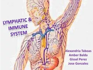
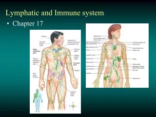
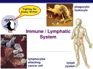
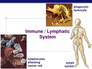
The lymphatic system • Functions • Return excess tissue fluid to bloodstream • Lacteals absorb fats • Defense against disease • Lymphatic vessels • One-way system • Begins with capillaries in tissues • Walls composed of simple squamous epithelium • Fluid inside is lymph • Water • Nutrients • Electrolytes • Cell products like hormones
Lymphatic capillaries merge to form vessels Vessels merge and empty into ducts Thoracic duct-empties into left subclavian vein from Body below thorax Left side of head Left arm Right lymphatic duct-empties into right subclavian vein from Right side of head Right arm
Vesselstructure • Small vessels • Simple squamous epithelium • Large vessels • Similar to veins • Valves to prevent backflow • Skeletal muscles “pump” lymph • Edema • Accumulation of tissue fluid • Occurs if not enough drainage, or too much produced • Can cause tissue damage and death
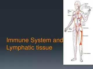
Lymphatic organs Contain lymphocytes Produced in thymus and bone marrow B lymphocytes-antibodies T lymphocytes-cellular immunity Primary lymphatic organs Red bone marrow Network of connective tissue fibers with sinuses Stem cells produce blood cells Most bones in children contain red marrow Adults- ends of long bones, skull, pelvis, clavicle, vertebrae B lymphocytes mature in red marrow
Primary lymphatic organs Thymus Between trachea and sternum above the heart Shrinks with age Divided into lobules by connective tissue Lobules filled with T lymphocytes Produced in bone marrow Mature in the thymus Produces hormones Thymosin-aids maturation of T lymphocytes
Secondary lymphatic organs Lymphocytes encounter antigens in secondary organs Lymphocytes then proliferate and active Spleen Upper left side of abdomen red pulp Vessels and sinuses Filters blood Macrophages destroy old RBC’s White pulp Clumps of lymphatic tissue Splenic capsule Thin, easily ruptured
Secondary lymphatic organs Lymph nodes Located along lymphatic vessels Divided into nodules by connective tissue Nodules packed with B and T lymphocytes Each contains a sinus Lymph filters through nodules Macrophages phagocytize pathogens and debris Lymph nodes named for location Inguinal nodes-groin Axillary nodes-armpit Cervical nodes-neck
Nonspecific and specific defenses • Nonspecific defenses • Barriers to entry • Skin and mucus membranes • Oil glands secrete antibacterial substances • Mucus • Acidic pH of stomach • Normal flora • Inflammatory reaction • Initiated by chemical agents or pathogens • 4 signs • Redness • Heat • Swelling • Pain
Inflammatory reaction • Signs due to capillary changes • Induced by chemical mediators • Histamine • Produced by mast cells • Vasodilator • Increases capillary permeability • Migration of phagocytes to damaged area • Neutrophils-from bloodstream • Monocytes-from bloodstream • Dendritic cells- in skin • Macrophages-in tissues • Pus- dead phagocytes and debris
Nonspecific defenses • Inflammation can be accompanied by other responses • Clot formation • Specific defenses mediated by T cells • Chronic inflammation • Persistent • Anti-inflammatory agents • Aspirin, NSAIDs, cortisone • Act against chemicals released by WBC’s in damaged area
Inflammatory reaction • Fig. 13.3
Nonspecific defenses • Natural killer cells • Large, granular lymphocytes • Kill virus infected cells and tumor cells • Kill by cell to cell contact • Detect antigenic changes in cancerous cells and infected cells • Nonspecific • Have no memory • Do not increase in number upon antigen exposure
Nonspecific defenses • Protective proteins • Complement system • Group of plasma proteins • Amplify inflammatory response • Bind to mast cells-stimulates histamine release • Attract phagocytes • Promote phagocytosis by binding to pathogens • Membrane attack complexes • Joined complement proteins • Produce holes in bacteria and viruses • Fluids and salts enter-lysis • Interferon • Produced by virus infected cells and binds to normal cells • Normal cells then release protective substances
Action of the complement system against a bacterium • Fig. 13.4
Specific defenses • Specific defenses • Function nonspecific have failed • Take 5-7 days to activate • Effects are long lasting • B cells and Antibody-mediated immunity • B cell receptor –BCR • BCR binds to specific antigen in lymph node or spleen • B cell then divides many times by mitosis-clone • Clones become • Plasma cells- produce antibodies to specific antigen • Memory cells-remember antigen for later exposure
Clonal selection theory as it applies to B cells • Fig. 13.5
Specific defenses • B cells and Antibody-mediated immunity. • Antibody structure • Y-shaped molecules with 2 “arms” • Each arm has a heavy and a light polypeptide chain • Constant region- specific for antibody type • Variable region-varies between antibodies • Hypervariable region- at tips of arms • Variable and hypervariable regions-antigen binding site • Lock and key fit • Antigen-antibody binding • Forms a complex • Marks antigen for destruction
Antibody structure • Fig. 13.6
Specific defenses • Table 13.1
Specific defenses • T cells and cell-mediated immunity • TCR- T cell receptor • Cannot recognize antigen without help • APC- antigen-presenting cell • Dendritic cells or macrophages • Phagocytize pathogen first • Break pathogens down • Display a piece of pathogen in major histocompatability complex (MHC) • On surface of APC membrane • Travel to nodes • “Present” antigen to T lymphocytes
Specific defenses • T cells and cell-mediated immunity cont’d. • T cell with specific TCR bind to antigen on macrophage surface • T cell becomes activated • Undergoes clonal expansion • Type of T cell formed depends upon MHC • MHC I- T cells formed are cytotoxic T cells • MHC II- T cells formed are helper T cells • Small number of clonal cells become memory cells • When infection clears, T cells undergo apoptosis
Clonal selection theory as it applies to T cells • Fig. 13.7
Specific defenses cont’d. • T cells and cell-mediated immunity • Types of T cells • Cytotoxic Tcells-cell mediated- immunity • Contain granules of perforins or granzymes • Cytotoxic T cell binds to pathogen and releases perforin • Perforin or granzyme enters cell and destroys it • Cell can move on to another target cell • Helper T cells • Secrete cytokines which activate all immune cells • Helper T cells needed for B cell activation • Helper T cells are infected by HIV virus
Cell-mediated immunity • Fig. 13.8
Induced immunity • Immunity • Naturally through exposure • Artificially (induced) through vaccination • Activeimmunity-individual produces antibodies against antigen • Passiveimmunity-individual is given prepared antibodies by injection • Active immunity • Immunization-use of vaccines to establish immunity • Vaccines-killed or attenuated pathogens
Induced immunity • Active immunity • Response to vaccination • Monitored by antibody titre • After first exposure, get primary response • First few days, no detectable antibodies • Then slow rise in number which levels off • Gradual decline as apoptosis occurs • Booster response • Boosts titre to high level • Rapid rise in antibody level • Prevents disease symptoms on subsequent exposures • Active immunity depends on memory T and B cells
Active immunity due to immunizations • Fig. 13.9
Induced immunity • Passive immunity • From prepared antibodies • Not produced by individual’s body • Temporary immunity • No memory cells formed • Used to prevent illness • Cytokines and immunity • Signaling molecules produced by T lymphocytes and macrophages • Interleukins • Cytokinins that enhance ability of T cells to fight cancer • May have many potential uses in medicine
Passive immunity • Fig. 13.10
Induced immunity • Monoclonal antibodies • Group of plasma cells from the same B cell all produce same antibody • Production of monoclonal antibodies • Mouse B lymphocytes exposed to antigen • Resulting cells fused with myeloma cells-hybridomas • Use of monoclonal antibodies • Diagnostic tests • Ex: pregnancy tests • Vehicles for drug delivery • Identification of infections
Production of monoclonal antibodies • Fig. 13.11
Immunity side effects • Allergies • Hypersensitivities to allergens • Immediate allergic response • Occurs within seconds • Mediated by IgE • Binding with antigen causes release of histamine from mast cells • Hay fever- reaction occurs in mucous membranes of nose and eyes • Asthma- reaction occurs in small airways • Anaphylactic shock • Can occur when allergen enters bloodstream • Life-threatening decrease in blood pressure from increased capillary permeability • Epinephrine can delay reaction
Allergies • Delayed allergic response • Initiated by helper T cells at site of allergen contact • Regulated by cytokines from macrophages and T cells • Ex: TB skin test • Tissue becomes red and hardened • Blood-type reactions • ABO system • Based on presence or absence of A and B antigens on RBC’s • If A present- blood is type A • If B present- blood is type B • If both resent- blood is type AB • If neither is present- blood is type O
Blood transfusions • Fig. 13.12
Blood-type reactions Rh system Another RBC antigen Antigen present- Rh positive Antigen absent- Rh negative Significant in pregnancy Rh neg mom pregnant with Rh pos baby If baby’s cells leak into mother’s bloodstream, she forms anti-Rh antibodies Attack baby’s RBC’s- hemolytic disease of newborn (HDN) Can affect subsequent Rh pos pregnancies as well Prevent by giving Rh neg mom anti-Rh immunoglobulins in an injection Must give BEFORE she becomes ensitized to produce her own
Hemolytic disease of the newborn • Fig. 13.13
Tissue rejection • Cytotoxic T cells recognize foreign antigens on transplanted organ or tissue • Transplanted organ is destroyed • Controlled by immunosuppressive drugs • Act by suppressing cytokines • Best success attained when MHC antigens of donor and recipient are closely matched • Xerotransplantation • Using organs of another species for transplantation • Pig is most commonly used-prolific, widely available • Genetic engineering can make pig organs less antigenic
Diseases of the immune system • Autoimmune diseases • Cytotoxic T cells or antibodies attack body’s cells • No cures available; controlled with drugs • Myasthenia gravis- neuromuscular junctions do not work • Multiple sclerosis-myelin sheath of neurons break down • Systemic lupus erythematosis- many systemic signs • Rheumatoid arthritis- affects joints • Immunodeficiency diseases • Immune system unable to protect against disease • Can be congenital from defect in lymphocyte formation • Can be infectious- HIV • Severe combined immunodeficiency disease-both T cells and B cells affected