Download
1 / 49
500 likes | 662 Views
ONTARIO. QUIT. BASE HOSPITAL GROUP. ADVANCED ASSESSMENT Cardiovascular System. 2007 Ontario Base Hospital Group. ADVANCED ASSESSMENT Cardiovascular System. AUTHORS Mike Muir AEMCA, ACP, BHSc Paramedic Program Manager Grey-Bruce-Huron Paramedic Base Hospital

E N D
ONTARIO QUIT BASE HOSPITAL GROUP ADVANCED ASSESSMENT Cardiovascular System 2007 Ontario Base Hospital Group
ADVANCED ASSESSMENT Cardiovascular System AUTHORS Mike Muir AEMCA, ACP, BHSc Paramedic Program Manager Grey-Bruce-Huron Paramedic Base Hospital Grey Bruce Health Services, Owen Sound Kevin McNab AEMCA, ACP Quality Assurance Manager Huron County EMS References – Emergency Medicine REVIEWERS/CONTRIBUTORS Rob Theriault EMCA, RCT(Adv.), CCP(F) Peel Region Base Hospital Donna L. Smith AEMCA, ACP Hamilton Base Hospital Tim Dodd, AEMCA, ACP Hamilton Base Hospital 2007 Ontario Base Hospital Group
CONSISTS OF: Heart (pump) Arteries and veins (container) Capillaries (site nutrient, gas exchange) Cardiovascular System
Transportation of oxygen and other nutrients to the cells Removal of carbon dioxide and wastes Distributes hormones Control heat transfer Functions
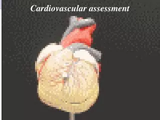
Left Ventricle High Pressure More Muscle Systemic Right Ventricle Low Pressure Less Muscle Pulmonary Heart Anatomy
Three Layers Endocardium Myocardium Epicardium Heart Anatomy
Automaticity All myocardial cells can generate an electrical impulse Conductivity Intercalated discs Contractility Functional syncitium Heart Physiology
Blood Flow Right atria via vena cava Tricuspid valve into right ventricle a) Pulmonic valve to pulmonary artery b) Right and left pulmonary arteries
Pulmonary arterioles to capillaries = gas exchange Blood Flow
Blood Flow Left atrium via pulmonary veins Mitral valve to left ventricle Aortic valve to aorta
Arteries Arterioles Capillaries Venules Veins Arteries & Veins
Three Layers Intima Media Adventitia Arteries & Veins
Left Main Left Anterior descending Circumflex Right RCA Marginal Posterior Decending Coronary Arteries
Lead Groups I aVR V1 V4 II aVL V2 V5 III aVF V3 V6 Limb Leads Chest Leads
Contractility Conductivity Automaticity Neuromuscular Electrophysiology
Contractility • Contractility • Similar to skeletal muscle • Interwoven muscle fibers
Thin Filament Actin Molecule Troponin Tropomyacin Muscle Fiber Contractility
Ca2+ = • Troponin = Muscle Contraction Contractility • Tropomyocin =
Cardiac versus Muscular Contractility
Specialized tissues conduct electrical impulses SA Node Intra-atrial pathways AV Node Bundle of His Lt and Rt Bundle Branches Purkinge Fibers Conductivity
mV 0 + 20 1 PURKINJE FIBRE 2 0 3 THRESHOLD POTENTIAL -65 4 4 -85 Na + INSIDE CELL OUTSIDE CELL - 100 - + + + + + + + + K Na Cl Ca K K K K Action Potential Phase 0: Rapid Depolarization Phase 3: Relative Refractory Period Phase 1: Early Repolarization Phase 4: Resting Membrane Potential Phase 2: Plateau ( Absolute Refractory Period)
Inherent ability of all myocardial cells to spontaneously depolarize Primary Pacemaker - SA Node Secondary – AV Node, Bundle of His, Bundle Branches, Purkinge Fibers Under stress all other cells can generate an impulse Automaticity
2 2 2 + 20 0 0 0 0 3 3 3 - 65 4 4 4 - 85 - 100 PACEMAKER CELL SA Node Phase 0: Depolarization Phase 3: Relative Refractory Period Phase 1: Does not Apply Phase 4: Spontaneous Phase 4 Rise Phase 2: Plateau ( Absolute Refractory Period)
Cardiac Output = Heart Rate x Stroke Volume Cardiac Output
Intrinsic Preload Extrinsic ANS Electrolytes Temperature Humoral/Chemical Cardiac Function Control
Preload Venous return to Heart 70 % blood volume Low Pressure Intrinsic
Autonomic Nervous System Extrinsic influences on CO
Electrolytes K+ - Increase will decrease rate and force Na+ - Increase will decrease force Ca++ - Increase will increase force Temperature Low - Decreased rate Hi - Increased rate,Increased force Humoral/Chemical Catecholamines – increase rate and force ADH – increased secretion increases preload Acids – increases in acids decreases function Extrinsic influences on CO
Rapid ANS Baroreceptors Chemoreceptors Blood Pressure Control
Blood Pressure Control • Intermediate • Renin/Angiotensin • ADH • Slow • Kidneys
Lub closing of A-V valves S1 Dub Closing of aortic and Pulmonic valves S2 Heart Sounds “lub dub”
When the heart is unable to pump the volume it receives it is said to be in failure Right Sided Left Sided Heart Failure
Causes Pump Failure Heart Failure
Causes Cardiac ischemia Hypertensive event Rate related Tachycardia Bradycardia Valvular disease Prolapse Rupture Heart Failure
Acute Right Sided Failure associated with acute inferior wall MI hypotension normal to slow heart rate JVD chest clear Treatment: fluid resuscitation Heart Failure Note: NTG contraindicated for HR < 60 and/or hypotension
Volume overload inappropriate fluid resuscitation diligent monitoring of respiratory status required when administering IV fluids Heart Failure Note: Auscultate chest q 250 cc in adults - q 100 cc in Paeds
Left or Right sidedheart failure Forward or Backward ventricular failure Backward failure is secondary to elevated systemic venous pressures. Forward ventricular failure is secondary to left ventricle failure and reduced flow into the aorta and systemic circulation CATEGORIZING FAILURE
Decreased emptying of the left ventricle Increased volume and end-diastolic pressure in the left ventricle Increased volume (pressure) in the left atrium Increased volume in pulmonary veins LV BACKWARDEFFECTS
LV BACKWARDEFFECTS cont’d. Increased volume in pulmonary capillary bed = increased hydrostatic pressure Transudation of fluid from capillaries to alveoli Rapid filling of alveolar spaces Pulmonary edema
Decreased cardiac output Decreased perfusion of tissues of body Decreased blood flow to kidneys and glands Increased reabsorption of sodium and water and vasoconstriction LV FORWARDEFFECTS
LVFORWARDEFFECTS cont’d. Increased secretion of sodium and water-retaining hormones Increased extracellular fluid volume Increased total blood volume and increased systemic blood pressure
Decreased emptying of the right ventricle Increased volume and end-diastolic pressure in the right ventricle Increased volume (pressure) in right atrium Increased volume and pressure in the great veins RV BACKWARDEFFECTS
RV BACKWARDEFFECTS cont’d. Increased volume in the systemic venous circulation Increased volume in distensible organs (hepatomegaly, splenomegaly) Increased pressures at capillary line Peripheral, dependant edema and serous infusion
Decreased volume from the RV to the lungs Decreased return to the left atrium and subsequent decreased cardiac output All the forward effects of left heart failure RVFORWARDEFFECTS
ONTARIO START QUIT BASE HOSPITAL GROUP Well Done! Ontario Base Hospital GroupSelf-directed Education Program