Download
1 / 79
800 likes | 997 Views
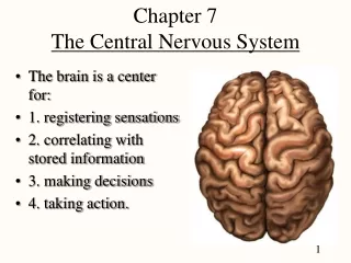
Chapter 7 The Nervous System. Biology 112 Tri-County Technical College Pendleton, SC. Structural Organization. Structural Classification which includes ALL nervous system organs has two subdivisions: CNS-consists of brain and spinal cord

E N D
Chapter 7 The Nervous System Biology 112 Tri-County Technical College Pendleton, SC
Structural Organization • Structural Classification which includes ALL nervous system organs has two subdivisions: • CNS-consists of brain and spinal cord • PNS(peripheral)-is part outside CNS consisting mainly of nerves that from CNS • Spinal nerves carry info to and from spinal cord • Cranial nerves carry info to and from brain
Functional Organization • Concerned ONLY with PNS structures • Sensory/Afferent division conducts impulses to CNS • Sensory fibers from skin, skeletal muscles, and joints are called somatic sensory (afferent) fibers • SFs from visceral organs called visceral sensory fibers (visceral afferents) • Sensory division keeps CNS informed of events going on outside/inside body
Functional Organization, cont. • Motor/Efferent division carries impulses from CNS to effector organs, muscles, and glands • Impulses activate muscles/glands • They EFFECT (bring about) a motor response • MOTOR division has two subdivisions • SOMATIC NERVOUS SYSTEM • AUTONOMIC NERVOUS SYSTEM
Autonomic Nervous System • ANS is motor subdivision of PNS that controls body activities automatically (involuntary) • Composed of special group of neurons that regulate cardiac muscle, smooth muscle, and glands • ANS also called involuntary nervous system • ANS has two distinct subdivisions • Sympathetic and Parasympathetic • Serve same organs but cause essentially opposite effects • Counterbalance each other to keep body systems running smoothly
ANS, continued • Preganglionic axons of sympathetic div. release acetylcholine; Postganglionic fibers release noreeinephrine and/or epinephrine (adrenergic fibers) • Pre- and Postganglionic axons of parasympathetic division release acetylcholine (cholinergic fibers) • Sympathetic part mobilizes body during extreme situations (fear, exercise, rage) • E for exercise/emergency/embarrassment • Parasympathetic part allows body to “unwind” and conserve energy • D for digesting/defecation/diuresis
Somatic Motor Division • Somatic system allows conscious/voluntary control of skeletal muscles • Often called “voluntary nervous system” • Cell bodies of motor neurons are inside CNS and their axons (in spinal nerves) extend all way to skeletal muscles served • Skeletal muscles are effectors of somatic nervous system • Neurotransmitter is acetylcholine
Cells of Nerve Tissue • Two principal types of cells • Neurons and supporting cells • Supporting cells of CNS lumped together as neuroglia (nerve glue) • Neuroglia (Glia) support, insulate, and protect delicate neurons • There are different types of “supporting cells” in the CNS
Supporting Cells, cont. • Astrocytes account for nearly ½ of neural tissue • Brace/anchor neurons to blood capillaries • Form living barrier between capillaries and neurons & play role in making exchanges between the two • Help protect neurons from harmful stuff in blood • Help control chemical environment by picking up excess ions and recapturing released neurotransmitters
Supporting Cells, cont. • Microglia are phagocytes that dispose of debris-dead brain cells, bacteria, etc. • Ependymal Cells line cavities of brain and spinal cord • beating of their cilia help circulate cerebrospinal fluid that fills those cavities and forms protective cushion around CNS • Oligodendrocytes wrap their flat extensions around nerve fibers producing fatty insulating coverings called myelin sheaths
FYI • Neuroglia are NOT able to transmit nerve impulses and they NEVER lose their ability to divide • Most brain tumors are GLIOMAS or tumors formed by glial cells
He ain’t heavy…he’s my brother • Supporting cells in PNS come in two major varieties • Schwann cells form myelin sheaths around nerve fibers that are found in PNS • Satellite cells act as protective, cushioning cells
You’re getting on my nerve.. • Neurons are highly specialized to carry impulses (transmit messages) • All neurons have CELL BODY (SOMA) and one or more slender processes extending from cell body • Dendrites conduct electrical impulses TOWARD the cell body • Axons conduct electrical impulses AWAY from cell body • May be 100s of dendrites but only 1 axon • Axons may branch at terminal end forming 100s to 1000s of axonal terminals
Neuron Part Functions • Cell body = nucleus and metabolic center • Dendrites = impulses to the cell body • Axon = impulses away from cell body to either another neuron or the effector • Most long neurons covered with whitish, fatty material called MYELIN • Myelin protects and insulates fiber and > the transmission rate of nerve impulses
Parts, cont. • Axons outside CNS insulated by Schwann cells • wrap tightly around axon jelly-roll style • wrapping done, tight coil of wrapped membranes called myelin sheath encloses axon • most of Schwann cell cytoplasm ends up just beneath outermost part of its plasma membrane • this part of Schwann cell, external to myelin sheath is called the neurilemma
Parts, cont. • Myelin sheath formed by many individual Schwann cells • Gaps or indentations called NODES OF RANVIER occur at regular intervals • Myelin sheaths in CNS are formed by the oligodendrocytes • CNS myelin sheath lacks a neurilemma • Neurilemma plays vital role in fiber regeneration of injured fiber • Regeneration of damaged fibers largely lacking in CNS
Nuclei, Ganglia, and more… • In CNS, cell bodies found in clusters called nuclei • This well protected location essential to well-being of nervous tissue • Small collections of cell bodies called ganglia are found outside CNS in the PNS • Bundles of nerve fibers running through CNS are called tracts • Bundles of nerve fibers running though PNS are called nerves
Nuclei and Ganglia, cont. • Terms white matter and gray matter refer to myelinated versus unmyelinated reions of the CNS • As a general rule, white matter consists of dense collections of myelinated fibers (tracts) • Gray matter contains mostly unmyelinated fibers and cell bodies
Classification of Neurons • Can be classified according to HOW they function or according to their STRUCTURE • FUNCTIONAL classification based on direction impulse traveling relative to CNS • Sensory (afferent) neurons carry impulses from sensory receptors (internal organs/skin) to CNS • Cell bodies of sensory neurons always found in ganglion outside the CNS • Inform about what is happening in/out of body
Classification, cont. • Motor neurons (efferent) carry impulses away from CNS to viscera/muscles/glands • Cell bodies of motor neurons always located in CNS • Association (interneurons) neurons connect sensory and motor neurons in a neural pathway • Cell bodies of association neurons always located in CNS
Structural Classification • Based on number of processes extending from cell body • Multipolar neuron has several processes • all motor and association neurons are multipolar • most common structural type • Bipolar neuron has 2 processes—dendrite and axon • Rare in adults; found only in some special sense organs (eye, ear) where act as sensory receptor cells
Structural Classification, cont. • Unipolar neuron has single process emerging from cell body • Very short and divides into proximal (central) and distal (peripheral) fibers • Unique in that only small branches at end of peripheral process are dendrites • Remainder of peripheral process and central process function as axon • In unipolar neuron, axon conducts impulse both toward and away from cell body • Sensory neurons found in PNS ganglia are unipolar
Resting State…I wish • Plasma membrane of resting (inactive) neuron is polarized • fewer positive charges on inner surface of membrane that on its outer face in tissue fluid • Major positive ions on inside are K+ • Major positive ions on outside are Na+ • As long as inside remains more negative as compared to outside, neuron will stay inactive (resting) • Resting potential of neuron is about –70 millivolts
Action, cameras, and more… • Action potential = nerve impulse • Stimulus changes permeability of patch of membrane and sodium ions diffuse rapidly into cell • Changes polarity of membrane at that location • Inside becomes more +, outside more – • Event called depolarization • If stimulus strong enough (at or > threshold) action potential is initiated
Action Potential, cont. • Depolarization of first membrane patch causes permeability changes in adjacent membrane and event is repeated • Membrane potential goes from –70 mv to +30 mv • Action potential propagates rapidly along entire length of membrane • Don’t want to waste space on this slide..so, IS THIS FUN, OR WHAT????
Repolarization • After patch of membrane depolarizes, it repolarizes • Membrane permeability changes and K+ ions diffuse OUT of cell • Restores negative charge inside cell and positive charge outside • Sodium-potassium pump used to restore ionic conditions of resting neuron • Resting potential of –70 mv restored • Until repolarization occurs, neuron CANNOT conduct another impulse
Reflex Defined and More… • Reflex is rapid, predictable and involuntary response to a stimulus • Reflexes occur over neural pathway called reflex arc • SOMATIC = reflexes that stimulate skeletal muscles • Dendrite of sensory neuron carries impulse to CNS • Processing of info may/may not occur in CNS • Axon of motor neuron carries impulse to effector • pull hand away from hot object = somatic reflex
Reflex, cont. • Autonomic reflexes regulate activity of smooth muscles, heart, and glands • saliva secretion and size of eye pupils two examples of autonomic reflexes • ALL reflex arcs have minimum of 5 elements • Sensory receptor which reacts to stimulus • Afferent neuron • Integration center located in CNS • Efferent neuron • Effector organ (muscle or gland to be stimulated) • Two-neuron (patellar knee-jerk) most simple • Three-neuron (flexor/withdrawal) more complex
Somatic reflexes • Patellar knee-jerk somatic reflex • receptors in patellar tendon • effectors are upper leg muscles that cause leg extension • Withdrawal somatic reflex • receptors located in epidermis/dermis • touching a hot object or finger stick • effectors are the appropriate muscles • can be used as a diagnostic tool • Whenever reflexes are exaggerated, distorted, or absent, nervous system disorders indicated
Cerebrum Functions • Speech • Memory • Logic • Emotional Responses • Consciousness • Interpretation of sensation • Voluntary movements
Basal Nuclei (ganglion) • Modify instructions from cerebrum to skeletal muscles • Problems with basal nuclei lead to inability to carry out movements in normal way • Huntington’s disease=inability to control muscles; individual exhibits abrupt, jerky, almost continuous movements • helped by drugs that block dopamine’s effect • Parkinson’s disease=trouble initiating movement or in getting muscles going • Persistent hand tremor with thumb and index finger making continuous circles with one another • Due to deficit of neurotransmitter dopamine
Functions of the Thalamus • Thalamus is part of diencephalon (innerbrain) that sits atop brain stem & enclosed by cerebral hemispheres • Encloses third ventricle of the brain • Relay station for sensory impulses > cerebrum • Provides “crude” recognition of whether sensation about to have is pleasant or unpleasant • Actual interpretation occurs in neurons of cerebral cortex
Functions of the Hypothalamus • Part of the diencephalon & important autonomic nervous system center • Plays role in regulation of body temp, water balance, and metabolism • Center for many drives and emotions • Important part of limbic system (emotional-visceral brain) • Thirst, appetite, sex, pain, and pleasure centers located in hypothalamus • Regulates pituitary gland and produces two hormones: ADH and oxytocin
Functions of the Midbrain • Midbrain is small part of the brain stem • Anteriorly composed of two bulging fiber tracts called cerebral peduncles that convey > and < impulses • Dorsally composed of four rounded protrusions called corpora quadrigemina which are reflex centers involved with vision and hearing
Functions of the Pons • PONS is rounded structure that protrudes just below midbrain • Functions as “bridge” for ascending and descending impulses through this area • Contains important nuclei involved with BREATHING
Functions of Medulla Oblongata • Most inferior part of the brain stem • Merges into spinal cord (no obvious change in structure) • Like pons, is important fiber tract area • Contains many nuclei that regulate vital visceral activities • Heart rate, blood pressure, breathing, swallowing, and vomiting
Functions of the Cerebellum • Has two hemispheres & convoluted surface • Outer cortex of gray matter and inner region of white matter • Provides precise timing for skeletal muscle activity, controls balance and equilibrium • It’s activity = smooth and coordinated body movements • Continually monitors brain’s intentions with action body performance by monitoring body position and amount of tension in various body parts
Protection for the CNS • CNS protected by enclosure within bone (skull and vertebral column); by watery cushion (cerebrospinal fluid) and by enclosure within membranes (meninges) • Also protected from harmful substances in blood by the blood-brain barrier • Three connective tissue membranes covering and protecting CNS structures are called meninges
CNS protection, cont. • Dura mater is leathery outermost layer • double layer membrane where surround brain • periosteal layer attached to inner surface of brain • meningeal layer forms outermost covering of brain and continues as dura mater of spinal cord • Arachnoid mater is weblike middle layer • Its threadlike extensions span subarachnoid space to attach it to innermost membrane, the pia mater • Pia mater (gentle mother) clings tightly to surface of brain and spinal cord by following every fold
CNS protection, cont. • Meningitis is inflammation of the meninges and is serious threat to brain because bacterial/viral meningitis may spread to nervous tissue of CNS • Meningitis usually diagnosed by taking sample of CSF from subarachnoid space • Brain inflammation called encephalitis
Cerebrospinal Fluid • CSF continually formed from blood by choroid plexuses (clusters of capillaries that hang from roof in each brain ventricle • Forms watery cushion in and around brain and spinal cord • Moves from lateral hemispheres 3rd ventriclecerebral aqueduct of midbrain 4th ventricle dorsalpons/medulla • Some fluid reaching 4th ventricle continues down central spinal canal of spinal cord