Download
1 / 54
670 likes | 864 Views
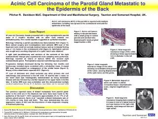
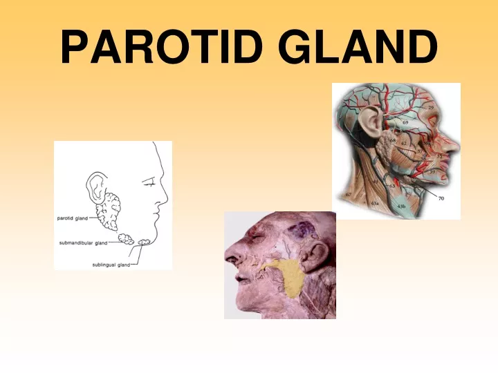
PAROTID GLAND. Parotid region. Facial lined space which forms the bed for the parotid gland Boundaries: In front – posterior border of ramus of mandible and medial pterygoid muscle behind – mastoid process and sternomastoid muscle Above – external acoustic meatus and TMJ

E N D
Parotid region • Facial lined space which forms the bed for the parotid gland Boundaries: • In front – posterior border of ramus of mandible and medial pterygoid muscle • behind – mastoid process and sternomastoid muscle • Above – external acoustic meatus and TMJ • Below – posterior belly of digastric • Medially – styloid process and styloid group of muscles
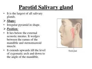
Parotid gland • Largest salivary gland • Inverted pyramid shape and is about 25 grams in weight • Lies below the external acoustic meatus
Coverings (capsule) • True capsule – by condensation of the stroma of the gland • False capsule – by splitting of investing layer of deep cervical fascia • The superficial lamina attached to the lower border of zygomatic arch • Deep lamina attached to the tympanic plate and styloid process • Deep lamina is thickened to form the stylomandibular ligament which separates the parotid from the submandibular gland
Presenting parts • Apex • Base • Three surfaces: -superficial - anteromedial - posteromedial • Three borders: - anterior - posterior - medial
Apex • Directed downwards and appears in carotid triangle • Overlaps posterior belly of digastric muscle • Structures passes through the apex: 1 cervical branch of facial nerve 2 anterior division of retromandibular vein
Base • Concave and directed above • Related to the external acoustic meatus and TMJ • Structures passes through the base: • Temporal branch of the facial nerve • Auriculo-temporal nerve • Superficial temporal vessels
Superficial surface • Covered by: • Skin • Superficial fascia • Platysma • Investing layer of deep cervical fascia • Great auricular nerve
Antero-medial surface • Deeply grooved for ramus of mandible • Related to: • Masseter • Posterior of ramus of mandible • Medial pterygoid muscle • Transmits branches of facial nerve and maxillary artery
Postero-medial surface • Relations: • Mastoid process and structures attached to it • Styloid apparatus • Internal carotid artery, internal jugular vein and last four cranial nerves • Facial nerve and external carotid artery pierces the gland through this surface
Anterior border • Thin and separates the superficial and anteromedial surfaces • Structures radiating from this border from above downwards: • Zygomatic branch of facial nerve • Transverse facial vessels • Upper buccal branch of facial nerve • Parotid duct • Lower buccal branch of facial nerve • Mandibular branch of facial nerve
Structures passing through the gland • From outside inwards: • Facial nerve • Retromandibular vein • External carotid artery
Parotid duct (stensen’s duct) • 5 cm in length • Emerges from the anterior border of gland • Runs forwards on the masseter muscle between upper and lower buccal branches • Turns medially at anterior border of masseter muscle • Pierces the buccal pad of fat, buccopharyngeal fascia and buccinator muscle • Passes obliquely between buccinator and mucous membrane of cheek • Opens in the vestibule of mouth on a papilla opposite the second upper molar tooth
Blood supply • Arterial – external carotid artery • Venous – external jugular vein
Nerve supply • Para sympathetic or secreto-motor pathway: preganglionic fibers arise from inferior salivatory nucleus tympanic branch of9th cranial nerve tympanic plexus lesser petrosal nerve otic ganglion
Nerve supply contd.. postganglionic fibers arises from the otic ganglion Auriculotemporal nerve Reaches to the gland • Effect of stimulation:watery secretion • Sympathetic nerve supply: • Plexus around the external carotid artery • Effect of stimulation:may reduce the secretion from the gland
Applied anatomy • Infection of parotid gland (mumps) • Abscess in the parotid gland • Siolography of the parotid duct • Accessory parotid gland • Frey’s syndrome
Submandibular salivary gland • Located in the digastric triangle • Size of a walnut • J-shaped • Indented by the posterior border of the mylohyoid • Divided into superficial and deep parts
Superficial part • Extends upto the mylohyoid line • Has inferior (superficial), medial and lateral surfaces • Partially enclosed between two layers of deep cervical fascia
Relations of superficial part • Inferior surface: skin, platysma, cervical br of facial nerve, deep fascia, facial vein, submandibular lymph nodes • Lateral surface: submandibular fossa, insertion of medial pterygoid muscle and facial artery • Medial surface: mylohyoid muscle, nerve and vessels, facial artery, hyoglossus, styloglossus, lingual nerve, submandibular ganglion and hypoglossal nerve
Deep part of gland • Lies deep to mylohyoid, superficial to hyoglossus and styloglossus • Continuous with the superficial part around the posterior border of the mylohyoid • Extends upto the sublingual gland
Submandibular duct • 5 cm long • Thin walled • Emerges from the deep part of submandibular gland • Runs forwards on the hyoglossus • Opens into the floor of the mouth • At the summit of the sublingual papilla on either side of the frenulum of the tongue
Blood supply & Lymphatic drainage • Facial artery • Common facial vein • Submandibular group of lymph nodes
Nerve supply • Parasympathetic: pathway • Superior salivatory nucleus • Sensory root of facial nerve • Geniculate ganglion of facial nerve • Chorda tympani nerve • Lingual nerve • Submandibular ganglion • Post-ganglionic fibers
Nerve supply contd • Sympathetic: plexus around facial artery • Sensory: Lingual nerve
Sublingual gland • Smallest of the three salivary glands • Almond shaped • Lies above the mylohyoid • Below the mucosa of the floor of the mouth • In the sublingual fossa
Sublingual gland • Drained by 15 ducts • Open either directly into the mouth or through the submandibular duct • Blood supply: Lingual and submental arteries • Nerve supply: similar to submandibular gland
Tempormandibular joint • Type : synovial joint • Subtype : condylar • Articulating bones: head of the mandible and mandibular fossa of base of the skull • Synovial cavity divided into upper and lower parts by an intra articular disc
Articulating bones • Upper articular surface: - mandibular fossa of the temporal bone and articular tubercle of temporal bone • Lower articular surface: - by the head of the mandible • Articular surfaces are covered with white fibro cartilage
Articular disc • Made of fibrocartilage • Upper surface is concavoconvex • Lower surface is concave • Divides the joint cavity into meniscotemporal and meniscomandibular compartments
Ligaments • Fibrous capsule: - attached to the margins of the articular surfaces - inner aspect lined by synovial membrane - lateral part strengthened by lateral temporomandibular ligament • Lateral temporomandibular ligament: - above attached to the tubercle of the root of the zygoma - lower end to the lateral aspect of the neck of the mandible
Ligaments contd… • Sphenomandibular ligament: - attached above to the spine of sphenoid and below to the lingula of the mandible - developed from the 1st pharyngeal arch • Stylomandibular ligament: - extends from tip of styloid process to the angle of the mandible - formed by thickening of deep fascia
Movements at the TMJ • Protraction: articular disc and head of the mandible glides forwards - Muscles responsible: masseter, medial and lateral pterygoid muscles • Retraction: backward gliding of articular disc in upper compartment and head of mandible in lower compartment - Muscles responsible: posterior fibers of temporalis muscle • Protraction and retraction occur in upper compartment of synovial cavity
Movements contd…. • Depression (opening of the mouth): - the head of the mandible moves in the under-surface of the disc and followed by forward gliding of disc and the head of the mandible - Muscle responsible: lateral pterygoid and suprahyoid and infrahyoid muscles • Elevation (closing of the mouth): - Muscles responsible: temporalis, masseter and medial pterygoid
Movements contd… • Chewing movements (side to side ): - head of one side glides forwards along with disc as in protrusion - head of the opposite side rotates on a vertical axis - chin moves forward and to the side - alternate movements of this kind on the two sides result in the side to side movements - Muscles responsible: temporal of one side, pterygoids of opposite sides, and the masseter
Applied anatomy • Dislocation of the TMJ
Muscles of mastication • Four muscles • Involved in the process of mastication • All supplied by the branches of the mandibular division of trigeminal • All located in the temporal and infratemporal fossae • All develop from the mesoderm of the first pharyngeal arch
Muscles of mastication Temporalis • Takes origin from the floor of the temporal fossa and the deep surface of the temporal fascia • Is inserted to the tip, medial surface and anterior border of the ramus of the mandible • Supplied by the deep temporal branches of mandibular nerve • Elevates and retracts the mandible
Muscles of mastication Masseter • Takes origin from the inferior border and medial surface of the zygomatic arch and maxillary process of zygomatic bone • Is inserted to the angle and lateral surface of the ramus of the mandible • Supplied by the massetric branches of mandibular nerve • Elevates and protrudes the mandible
Muscles of mastication Lateral pterygoid • Takes origin from the (a) infratemporal surface and crest of the greater wing of the sphenoid and (b) lateral surface of the lateral pterygoid plate • Is inserted to the joint capsule and disc of TMJ, pterygoid fovea on the neck of the mandible • Supplied by the nerve to lateral pterygoid • Bilaterally, protracts and depresses; unilaterally, swings the jaw to the contralateral side