Download
1 / 28
290 likes | 407 Views
PAROTID GLAND. Dr Shivarama Bhat Curtsey: Slideshare. Salivary Glands : 3 paired – Parotid Submandibular Sublingual Scattered - Tongue Palate

E N D
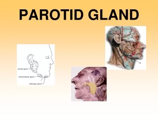
PAROTID GLAND Dr Shivarama Bhat Curtsey: Slideshare
Salivary Glands : 3 paired – Parotid Submandibular Sublingual Scattered - Tongue Palate Cheeks Lips Function : Chewing Swallowing Saliva (Digestion by Enzymes) Serous & Mucinous
GUT TUBE Stomodeum Buccopharyngeal membrane Pre-Laryngeal (Cephalic part) Pharynx and Part of definitive Mouth cavity Skin ectoderm Fore-gut Post-Laryngeal (Caudal part) Ectodermal furrow Mid-gut Parotid gland 4th week of IUL Hind-gut Proctodeum Cloacal membrane
The salivary glands arise as buds from The epithelial lining of the mouth; the parotid appears during the fourth week In the angle between the maxillary process and the mandibular arch Opening of parotid duct indicates position of angle of primitive mouth
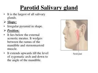
PAROTID SALIVARY GLAND
Parotid region Ext. acoustic meatus Sternomastoid Ramus of mandible
Parotid gland: Largest saliv. Gland Wt. 15 gms Serous secretion Capsule – True – condensation of fibrous stroma of gland False - investing deep fascia Superficial thick – adherent to gland Deep – thin – attached to styloid process forms – stylomandibular Lig. (separates parotid from submandibular sal. gland.
Parotid region Ext. acoustic meatus Sternomastoid Ramus of mandible
Parotid – 3 sided pyramid Medial border Antero-medial surface Postero-medial surface Superior surface (Base) Superficial surface Anterior border Posterior border Apex
Presenting parts • 01. Apex • 02. Base • 03. Superficial (lateral) surface • 04. Antero-medial surface • 05. Postero-medial surface
Apex • Overlaps post. Belly of digastric • appears in carotid triangle, related to • stylomandibular ligament • Structures passing thro’ • 01. Cervical branch of facial nerve • 02. Ant. division of retro-mandibular vein • 03. Formation of ext. jug. vein
2. Base Concave related to; Ext. acoustic meatus Post. Part of TM joint Structures passing thro’ 01. Temp. br. Of facial nerve 02. Sup. Temp. vessels 03. Auriculo-temp. nerve
3. Antero-medial surface Deeply grooved by ramus of mandible; Relations: 01. Post. Inf. Part of masseter 02. Post. Border of ramus of mandible 03. Med. Pterygoid muscle insertion 04. Outer lip – branches of facial nerve 05. Inner lip - Maxillary art. Medial to neck of mandible 05 03 05 01 02
3. Postero - medial surface • Extensive • Relations: • Mastoid process with Sternomastoid • Post. Belly of digastric • 02. Styloid process with muscles • 03. Transverse process of atlas and • Rectus capitis lateralis • 04. Deep to styloid – Int. carotid • - Int. Jug. Vein • in between • 9, 10, 11, 12 cr. nerves • 05. Facial nerve after emerging from • SM foramen pierces the gland • 06. Ext. carotid first lodges in groove then • pierces the gland
1. Anterior border From above downwards: 01. Zygomatic br. of facial nerve 02. Transverse facial vessels 03. Upper buccal br. of facial nerve 04. Acc. Parotid gland & duct if present 05. Parotid duct 06. Lower buccal br. of facial nerve 07. Marginal mandibular br. of facial nerve 2. Posterior border Rests on masseter 01. Post. Auricular br of facial nerve 02. Post. Auricular vessels
Parotid duct (stensen’s duct) 5 cms long, starts at mid point of ant. Border Runs on masseter, opens opp. crown of upper 2nd Molar Sup: Upper buccal br. of facial, Transverse facial vessels. Inf: Lower buccal br. of facial Pierces:1.Buccal pad of fat, 2. Buccopharyngeal fascia 3. Buccinator
Parotid duct Surface marking
Processes of the parotid gland: 01. Facial process -Superficial to masseter along parotid duct 02. Acc. Parotid gland and duct if present 03. Pterygoid process – Bet’ mandibular ramus and ptrygoid muscle 04. Glenoid process - Bet. Ext meatus and capsule of TM joint 05. Pre and post styloid processes in front related to int. carotid behind related to int. Jug. Vein
Structures passing through parotid • Facial nerve • Retromandibular vein • External carotid
Branching of facial nerve (Radiate like goose’s foot – Pes anserinus) Emerges from SM foramen Pierces post. Medial surface Lies superficial to Retromeandibualr vein & Ext. carotid art. Divides into 1. Temporo faicial division - Temporal Zygomatic 2. Cervico facial division - Buccal Marginal mandibular Cervical
Blood supply Arterial – branches of ext. carotid Veins - to ext. jugular vein Lymphatics – parotid nodes Nerve supply – Secretomotor from both Parasympathetic – Serous Inf. Salivatory nucleus - medulla (auriculo temporal) Sympathetic - Mucinous Sup. Cerv. ganglion (Ext. carotid art.) Vasomotor
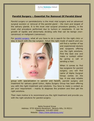
Applied anatomy Parotitis – ext capsule adherent – more painful Parotid abscess – horizontal incision Mixed parotid tumor – facial nerve not involved Frey’s syndrome – Penetrating wounds secretomotor fibres of auriculotemporal join with great auricular perspiration of skin covering parotid during eating