Download
1 / 21
290 likes | 1.42k Views
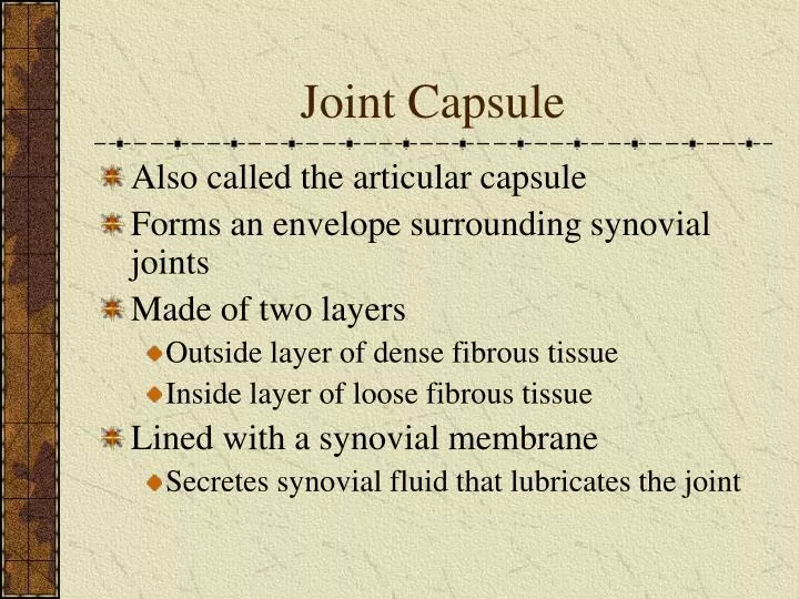
Joint Capsule. Also called the articular capsule Forms an envelope surrounding synovial joints Made of two layers Outside layer of dense fibrous tissue Inside layer of loose fibrous tissue Lined with a synovial membrane Secretes synovial fluid that lubricates the joint. Synovial Joint.

E N D
Joint Capsule Also called the articular capsule Forms an envelope surrounding synovial joints Made of two layers Outside layer of dense fibrous tissue Inside layer of loose fibrous tissue Lined with a synovial membrane Secretes synovial fluid that lubricates the joint
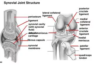
Synovial Joint joint capsule joint capsule articular cartilage
Joint Capsule (continued) • Help to hold bones together across synovial joints • Fairly tough and inelastic • Loose enough so as not to restrict normal joint movement
Shoulder Joint – Cross Section articular cartilage joint capsule humerus scapula labrum joint capsule
Ligaments • Made of dense fibrous tissue with the fibers in roughly parallel lines in the direction of functional need • In most cases pliable but inelastic • Bind bones to bones • Supply passive support and guidance to joints
Ligaments (continued) • May be so integrated into the joint capsule that they are indistinguishable as separate structures • Reinforce joint stability • Generally loose enough to allow free joint movement within a desirable range • Designed to prevent movement in a range which would be damaging
Ligaments (continued) • Major biochemical constituents • Water – 60% to 80% of wet weight • Collagen – 70% of the dry weight (mostly type I collagen) • elastin, glycoproteins, protein, polysaccharides, glycolipids, and cells (mostly fibroblasts) • Collagen turnover rate • Average of 300 to 500 days • Several months may be required to adapt, rebuild, or repair
Ligaments (continued) • Blood supply • Poor in the ligament itself • Ligaments depend on diffusion to supply inner fibers with nutrients • Vascular damage during injury can be particularly bad for the healing of ligaments • Blood flow is facilitated by passive movements after injury or surgery
Crimping • Collagen fibers in unstressed ligaments take on a wavy pattern • A result of collagen fibers cross-linking with elastic and reticular fibers • Straightening out of the crimp pattern produces the “toe” region of the ligament stress-strain curve
Crimping (continued) section of ligament collagen bundle crimp structure (tends to straighten out when the ligament undergoes tensile stress)
Ligament Stress-Strain Curve failure yield point plastic elastic toe
Viscoelastic Properties of Ligaments • Hysteresis – The loss of energy through the process of loading and then unloading • Ligaments undergo this energy loss because of internal friction during deformation • Relaxation (or Stress Relaxation) • Ligaments relax or lose tension if held in a stressed position for an extended period
Viscoelastic Properties of Ligaments • Load Elongation • Ligaments will tend to elongate or stretch under a constant tensile load when held for an extended period • Time Dependent Behavior of Ligaments (Ligament Softening) • Ligaments tend to relax or produce less reaction force to loads after extended athletic performance • In one study, knee laxity increased by more than 18% after 90 minutes of basketball or after a 10 k run
Sensory Receptors in Ligaments • May supply feedback to muscle tissue • Allow muscles to be recruited to assist in joint stability • A poorly understood phenomenon • Requires further investigation
Force Dissipation • Force is effectively dissipated at the ligament-bone junction • Failure is more likely to occur in the bone or ligament than at the junction
Failure Mechanisms • Ligamentous Failure – The ligament itself fails • Characteristic of fast loading rates • Bone Avulsion – The bone itself fractures or fails beneath the junction of ligament and bone • Characteristic of slow loading rates • Failure at the Ligament-Bone Junction • The least common type of failure
Effects of Aging • During Maturation • Structural and mechanical properties improve • Immediately After Reaching Full Maturity • Tissue strength may increase because of collagen stabilization • Loss of water and elastin reduces plasticity
Effects of Aging (continued) • After Maturity • Stiffness (resistance to deformation) decreases • Less deformation (strain) to failure • Decreased load to produce failure (strength decreases) • Bone avulsions increase at age 50 and above • Water content decreases resulting in reduced pliability • Elasticity is reduced
Ligamenta Flava (elastic ligaments) • Found between vertebrae • Contain yellow elastic tissue • Keep the vertebrae tightly against each other • There are inelastic ligaments between vertebrae that are relatively loose but prevent movements which would overstretch the ligamenta flava • Individual ligamenta flava are called ligamentum flavum
Vertebrae – Cross Section inelastic ligaments ligamentum flavum