Download
1 / 34
350 likes | 719 Views
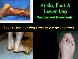
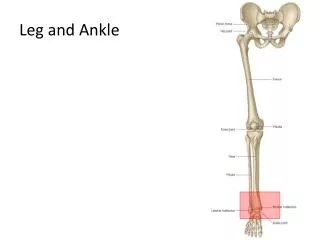
Leg, foot and ankle joint. MSK Revision SCRUBS Zahir Mughal. Introduction. Leg Osteology Musculature Neurovascular structures Clinical correlates Foot (brief) Ankle Anatomy Clinical correlates Pictures from Gray’s anatomy. Leg. Leg Osteology. Musculature of leg.

E N D
Leg, foot and ankle joint MSK Revision SCRUBS ZahirMughal
Introduction • Leg • Osteology • Musculature • Neurovascular structures • Clinical correlates • Foot (brief) • Ankle • Anatomy • Clinical correlates • Pictures from Gray’s anatomy
Muscles of the leg • Anterior compartment • Tibialis anterior • Extensor hallucislongus • Extensor digitorumlongus • Fibularistertius
Lateral compartment • Fibularislongus • Fibularisbrevis
Posterior compartment • Superficial group • Soleus • Gastrocnemius • Plantaris • Deep group • Popliteus • Flexor digitorumlongus • Tibialis posterior • Flexor hallucislongus
Actions of leg muscles • Anterior compartment • Dorsiflex the foot at the ankle joint • Extend the toes • Invert the foot • Lateral compartment • Evert the foot • Posterior compartment • Superficial group • Plantarflex the foot at the ankle joint • Deep group • Popliteus unlocks the extended knee • Plantarflex and invert the foot and flex the toes
Compartment syndrome • Raised intra-compartmental pressure • Intra-compartmental pressure > intra-arterial pressure • Arterial occlusion ischemia • Presentation 6Ps • 1. PAIN • 2. PAIN • 3. PAIN • 4. PAIN • 5. PAIN • 6. PAIN Medical Emergency
Arteries of leg • Popliteal artery is the major blood supply to the leg and foot. • Starts at the adductor hiatus • Traverses the poplitealfossa • Ends at inferior border of popliteus • Divides into anterior and posterior tibial arteries • Anterior compartment supplied by anterior tibial artery • Lateral compartment and posterior compartment supplied by posterior tibial artery
QUIZ - Arteries of leg Peroneal artery
Veins of leg • Deep veins follow arteries • Superficial veins • Long saphenous vein • Short saphenous vein
Varicose veins • Long, dilated, tortuous superficial veins • Incompetent valves • Sapheno-femoral junction • Sapheno-popliteal junction • Perforators • Risk factors • Prolonged standing, obesity, pregnancy, family history, oral contraceptive pill • Surgery
QUIZ – Veins of the leg Femoral vein • TRUE or FALSE • 1. Veins start proximally and end distally? • F • 2. Long saphenous vein is a branch of Femoral vein? • F • 3. Short saphenous vein is posterior to lateral malleolus? • T • 4. Superficial veins drain into deep veins via perforator veins? • T Popliteal vein Short saphenous vein Long saphenous vein Dorsal venous arch
Nerves of the leg • Sciatic nerve (L4-S3) divides into tibial nerve and common fibular nerve • Common fibular nerve divides into superficial and deep branches • Anterior compartment supplied by: Deep fibular nerve • Lateral compartment supplied by: Superficial fibular nerve • Posterior compartment supplied by: Tibial nerve
Nerve injury • Sciatic nerve • Loss of extension of thigh, flexion of knee, all motor function of foot and toes • Loss of sensation of lower leg and foot • Common peroneal nerve (bumper fractures) • Loss of extension of toes and foot (footdrop) • Loss of sensation of lateral lower leg and upper foot
PoplitealFossa • Boundaries • Superomedial – Semimembranosus/semitendinosus • Superolateral – Biceps femoris • Inferomedial – medial head of gastrocnemius • Inferolateral – lateral head of gastrocnemius and plantaris • Floor – femur, popliteus, capsule • Roof – popliteal fascia, skin • Contents – • Small saphenous vein • Popliteal artery and vein • Tibial and common fibular nerves • Posterior cutaneous nerve of thigh • Popliteal lymph nodes and lymphatic vessels
Poplitealfossa • Differentials for mass in the poplitealfossa • Lipoma • Bakers cyst • Popliteal aneurysm • Popliteal Abscess • Neoplasm • Lymphadenopathy
QUIZ - PoplitealFossa Medial Lateral
Arches of the foot • Absorb and distribute downward forces • Ligaments and muscles support the arches of the foot
Blood supply • Arterial: • Posterior tibial and dorsalispedis supply the foot • Venous: • Deep veins follow the arteries • Superficial veins arise from dorsal venous arch
Nerve supply • The foot is supplied by the (1) tibial, (2) deep peroneal, (3) superficial peroneal, (4)sural, and (5)saphenous nerves • all five nerves contribute to cutaneous or general sensory innervation • the tibial nerve innervates all intrinsic muscles of the foot except for the extensor digitorumbrevis, which is innervated by the deep fibular nerve
Ankle joint • Synovial joint • Movements: • hinge-like dorsiflexion and plantarflexion of the foot • Ankle is most stable when the foot is dorsiflexed • Strong ligaments stabilise the ankle joint
Subtalar joint • Articulation between: • inferior surface of the talus • superior surface of the calcaneus • Movement: • Inversion and eversion of the foot • Involved with inversion and eversion of the foot. • ( Talocalcaneonavicular and Calcaneocuboid joint joints )
Ligaments of ankle joint • Medial (deltoid) ligament • Large and very strong • Lateral ligament
Pott’s Fracture • Mechanism • Forced foot eversion • Deltoid ligament pulls on the medial malleolus and causes avulsion • Talus displaced laterally and posterior and causes avulsion of the lateral malleolus • Both malleoli are fractured (bimalleolar fracture)
Medial malleolus relations • Posterior: • Timothy Doth Vex All Nervous Housemaids (ML) • Tibialis posterior • Flexor digitorumlongus • Posterior tibial vein • Posterior tibial artery • Tibial nerve • Flexor hallucuslongus • Anterior: • Long saphenous vein • Saphenous nerve
Lateral malleolus relations • Posterior: • Short saphenous vein • Sural nerve • Anterior: • Peroneal artery
Conclusion • Anatomy of the leg • Compartments • Muscles • Vessels • Nerves • Clinical relevance • Anatomy of the foot and ankle joint • Brief overview • Questions?