Download
1 / 53
540 likes | 808 Views
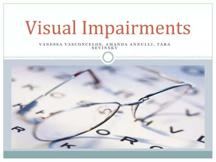
Visual Impairments. Vanessa Vasconcelos, amanda annulli , tara Sevinsky. Anatomy of the Eye. Retina Macula Fovea Rods Cones Optic Nerve. http://t2rerc.buffalo.edu/pubs/ip/VI/IP_VI.pdf. Retina. the innermost layer in the eye

E N D
Visual Impairments Vanessa Vasconcelos, amandaannulli, taraSevinsky
Anatomy of the Eye Retina Macula Fovea Rods Cones Optic Nerve http://t2rerc.buffalo.edu/pubs/ip/VI/IP_VI.pdf
Retina the innermost layer in the eye converts impulses that are sent along the optic nerve to the brain composed of light sensitive cells known as rods and cones the retina can be compared to the film of a camera http://t2rerc.buffalo.edu/pubs/ip/VI/IP_VI.pdf
Rods & Cones • Rods: • Cells that are primarily in the outer retina • Do not discriminate colors • Low spatial resolution • Supports vision in low light • “night vision” • Sensitive to object movement • Provide peripheral vision • Cones: • Cells that are densely packed within the central visual field • Function best in bright light • Process acute images • Discriminate colors http://t2rerc.buffalo.edu/pubs/ip/VI/IP_VI.pdf
Optic Nerve the second cranial nerve connects the eye to the brain carries impulses formed by the retina the nerve layer that lines the back of the eye and senses light and creates impulses the impulses are dispatched through the optic nerve to the brain, which interprets them as images http://t2rerc.buffalo.edu/pubs/ip/VI/IP_VI.pdf
Macula Located in the back of the eye In the center of the retina Within the macula is an area called the fovea http://t2rerc.buffalo.edu/pubs/ip/VI/IP_VI.pdf
Fovea A tiny pit located in the macula of the retina Contains the highest concentration of cones Produces the sharpest vision, and is used to see details clearly Only in the fovea are the layers of the retina spread aside to let light fall directly on the cones, the cells that give the sharpest image http://t2rerc.buffalo.edu/pubs/ip/VI/IP_VI.pdf
Visual Acuity • What is it? • Visual acuity is an indication of the clarity or clearness of one’s vision. • The measurement of how well a person sees. • What is considered “normal” vision? • 20/20 vision • They are able to see what someone with “normal” vision can see at 20 feet away. http://t2rerc.buffalo.edu/pubs/ip/VI/IP_VI.pdf
What is used for a standard eye exam? Visual acuity is tested by reading a Snellen eye chart at a distance of 20 feet The eye chart is imprinted with block letters that decrease in size line-by-line, corresponding to the distance at which that line of letters in normally visible http://t2rerc.buffalo.edu/pubs/ip/VI/IP_VI.pdf
Legal Blindness A level of vision loss that has been legally defined to determine eligibility for benefits Refers to a diagnosis of 20/200 or less in the better eye with the best possible correction and/or a visual field of less than 20 degrees Often people who are diagnosed with legal blindness still have some useable vision • http://www.afb.org/section.aspx?sectionid=15&documentid=1280
Total Blindness Considered to be present when there is no light perception Refers to an inability to see anything with either eye http://t2rerc.buffalo.edu/pubs/ip/VI/IP_VI.pdf
Cortical Blindness Also called cortical visual impairment (CVI) Total or partial loss of vision in a normal-appearing eye caused by damage to the brain’s occipital cortex A condition in which the eyes and optic nerves appear healthy; yet, the patient does not have normal vision or normal visual perception The eye itself is normal, but the brain does not process the information properly Can be temporary or permanent Cortical Visual Impairment. (n.d.). California School for the Blind. Retrieved November 20, 2012, from http://www.csb-cde.ca.gov/Documents/Causes%20of%20Visual%20Impairment/ causes_cortical_visual_impairment.htm
Causes • Traumatic brain injury • Head trauma • Seizures • Lack of oxygen before, during or after birth • Perinatal Hypoxia is the most common cause seen in children • Stroke • Viral or bacterial infection • Intracranial bleeding • ANY CONDITION THAT AFFECTS THE CORTEX OF THE BRAIN Cortical Visual Impairment. (n.d.). California School for the Blind. Retrieved November 20, 2012, from http://www.csb-cde.ca.gov/Documents/Causes%20of%20Visual%20Impairment/ causes_cortical_visual_impairment.htm
Characteristics of Cortical Blindness • Normal or minimally abnormal eye exam • Visually attends in near space only • Difficulties with visual complexity/crowding • Individual performs best when one sensory input is presented at a time, when the surrounding environment lacks clutter, and the object being presented is simple • Non-purposeful gaze/light gazing behaviors • Distinct color preference • Preferences are predominantly red and yellow, but could be any color • Visual field deficits • It is not so much the severity of the field loss, but where the field loss is located.) • Visual latency • The individual's visual responses are slow, often delayed • Attraction to movement, especially rapid movements. • Absent or atypical visual reflexive responses • Atypical visual motor behaviors • Momentary fixation – • (fixate) on things only briefly, • Avoidance – • Some children with CVI will actively avoid (e.g., look away from) salient visual objects Cortical Visual Impairment. (n.d.). Home Page. Retrieved November 20, 2012, from http://www.ohiolionseyeresearch.com/cortical
Associated Difficulties • Many have diagnoses such as cerebral palsy (about 80%), epilepsy, hydrocephalus, and deafness • Difficulty with color and depth perception • Very slow brain processing skills • Difficulty with figure-ground • Seeing an object and not just the background • Lighting may be an issue • Too bright, not bright enough • Unusual visual behavior • Like to use peripheral vision or sometimes other senses to identify an object • Inability to see object close together Cortical Visual Impairment. (n.d.). Home Page. Retrieved November 20, 2012, from http://www.ohiolionseyeresearch.com/cortical
Prevalence and Prognosis • Can occur at any age when injury/illness occurs • Most commonly seen in children who suffered from hypoxia (low or no oxygen) • Vision fluctuates over time • Can improve overtime or stay same depending on cause • Temporary/Mild CVI, caused by minor fall may begin to improve days or months after the incident • Permanent/Severe CVI, caused by perinatal hypoxia may slightly improve but usually never fully improves • *PROGNOSIS DEPENDS ON AGE ON ONSET, TYPE, AND SEVERITY* Cortical Visual Impairment. (n.d.). California School for the Blind. Retrieved November 20, 2012, from http://www.csb-cde.ca.gov/Documents/Causes%20of%20Visual%20Impairment/ causes_cortical_visual_impairment.htm
Refractive Disorders Myopia* Hyperopia* Presbyopia Astigmatism
Myopia Also known as “near sightedness” Results from elongation of the eyeball Causes the image to fall in front of he retina instead of on its surface Difficulty seeing objects at a distance, tasks that require near vision are unaffected (they are able to see close up but not far away) 1/3 of the American population is affected http://t2rerc.buffalo.edu/pubs/ip/VI/IP_VI.pdf
Hyperopia Also known as “far sightedness” Caused by a slightly shortened eyeball Causing images to focus slightly behind the retina Affects vision for close tasks ¼ of Americans have hyperopia http://t2rerc.buffalo.edu/pubs/ip/VI/IP_VI.pdf
Common treatments Corrective lenses or glasses 150 million Americans spend $15 billion per year on corrective lenses supporting a $30 billion optical industry Many Americans are beginning to turn to surgery to correct both myopia and hyperopia http://t2rerc.buffalo.edu/pubs/ip/VI/IP_VI.pdf
Age Related Eye Disorders • Retinal Disorders • Age related macular degeneration • Wet and dry • Diabetic retinopathy • Retinal detachment • Cataracts • Glaucoma: open-angle and angle-closure • Dry Eye • Low Vision • Presbyopia
Retinal Disorders Macular Degeneration Diabetic Retinopathy Retinal Detachment *All have issues with the retina of the eye
Age Related Macular Degeneration (AMD) • Chronic condition that causes central vision loss • Symptoms include • blurriness, wavy lines, or a blind spot. • Causes/ Risk factors • Things you CAN control: Smoking, high cholesterol, high blood pressure, obesity • Things you CANNOT control: age, family history, gender and race (more common in females and whites) Haddrill, M. (n.d.). Age-Related Macular Degeneration - A Complete Guide. All About Vision - Complete Consumer Guide About Vision and Eye Care. Retrieved November 22, 2012, from http://www.allaboutvision.com/conditions/amd.htm
AMD: Wet and Dry • Two types of AMD: Wet and Dry • Dry AMD • Less severe, more common • is an early stage of the disease and may result from the aging and thinning of macular tissues, depositing of pigment in the macula or a combination of the two processes. • diagnosed when yellowish spots known as drusen begin to accumulate in and around the macula. • It is believed these spots are deposits or debris from deteriorating tissue. • Wet AMD • More severe, less common • Marked by swelling caused by leaking blood vessels that affect the macula • Almost always begins as dry macular degeneration. • Unknown cause of wet AMD • Early detection and treatment may help reduce the extent of vision loss and improve vision Wet macular degeneration - MayoClinic.com. (n.d.). Mayo Clinic. Retrieved November 22, 2012, from http://www.mayoclinic.com/health/wet-macular-degeneration/DS01086
AMD Treatmentand Prevalence • Seen most commonly in older adults, becomes more prevalent with adults age 60 and older • No cure, but some treatments may delay its progression or even improve vision • Treatments for macular degeneration depend on whether the disease is in its early-stage, dry form or in the more advanced, wet form • Treatments such as medication, surgery, eye injections, and nutrition can help prevent and sometime improve AMD Macular Degeneration Treatments. (n.d.). American Health Assistance Foundation (AHAF): Alzheimer's disease, macular degeneration and glaucoma.. Retrieved November 20, 2012, from http://www.ahaf.org/macular/treatment/
Diabetic Retinopathy/Diabetic Macular Edema (DME) • Leaky blood vessels in the retina • Happens when these damaged blood vessels leak fluid into the macula, this causes the macula to swell • Prevalence • Occurs mostly in people who have diabetes • 40-45% of people with diabetes have some stage of DME • Both type 1 and type 2 diabetes at risk • Common with woman who are pregnant and have diabetes • Risk Factors • Having diabetes for more then 10 years, high blood sugar levels, high fasting glucose levels, high cholesterol, high blood pressure, smoking • Symptoms • Small patches of vision loss • Colors seem to be “washed out” or changed • Straight lines look bent or crooked About Diabetic Macular Edema (DME) - Lucentis® (ranibizumab injection). (n.d.). Lucentis® (ranibizumab injection) for Diabetic Macular Edema (DME) and Wet Age-Related Macular Degeneration (Wet AMD).. Retrieved November 22, 2012, from http://www.lucentis.com/lucentis/dme/about_dme.html
DME: Stages • Mild NonproliferativeRetinopathy • Microaneurysms occur • small areas of balloon-like swelling in the retina's tiny blood vessels • Moderate NonproliferativeRetinopathy • Disease progresses, some blood vessels that nourish the retina are blocked • Severe NonproliferativeRetinopathy • Many more blood vessels are blocked, depriving several areas of the retina with their blood supply • These areas of the retina send signals to the body to grow new blood vessels for nourishment. • Proliferative Retinopathy • the signals sent by the retina for nourishment trigger the growth of new blood vessels. • This is called proliferative retinopathy • These new blood vessels are abnormal and fragile • They grow along the retina and along the surface of the clear, vitreous gel that fills the inside of the eye. • these blood vessels do not cause symptoms or vision loss. • However, they have thin, fragile walls, if they leak blood severe vision loss and even blindness can occur Diabetic Eye Disease, Facts About [NEI Health Information]. (n.d.). NEI | National Eye Institute Homepage. Retrieved November 22, 2012, from http://www.nei.nih.gov/health/diabetic/retinopathy
DME: Treatment • During the first three stages of diabetic retinopathy, no treatment is needed, unless you have macular edema • Macular edema is treated with a focal laser procedure • Proliferative retinopathy (4th stage) is treated with laser surgery • This procedure is called scatter laser treatment. • Scatter laser treatment helps to shrink the abnormal blood vessels. • If the bleeding is severe, you may need a surgical procedure called a vitrectomy. • Blood is removed from the center of your eye. Diabetic Eye Disease, Facts About [NEI Health Information]. (n.d.). NEI | National Eye Institute Homepage. Retrieved November 22, 2012, from http://www.nei.nih.gov/health/diabetic/retinopathy
Retinal Detachment • A separation of the retina from its attachments to the underlying tissue within the eye • Prevalence • Retinal detachments can occur at any age • occur most commonly in younger adults (25-50 years of age) who are highly nearsighted (myopic) and in older people following cataract surgery • Symptoms • Flashing lights and floaters may be the initial symptoms • Shadow or curtain that affects any part of the vision can indicate that a retinal tear has progressed to a detached retina. • Peripheral vision is lost first, progress leads to full vision loss • Caused by a retinal break, hole, or tear • Vitreous gel pulls loose or separates from the retina • Injury, age, surgery • Risk Factors • Cataract surgery, family history, diabetes Retinal Disorders: MedlinePlus. (n.d.). National Library of Medicine - National Institutes of Health. Retrieved November 20, 2012, from http://www.nlm.nih.gov/medlineplus/retinald
Retinal Detachment:Treatment • Treatment • Early diagnosis and repair are urgent since visual improvement is much greater when the retina is repaired before the macula or central area is detached. • Retinal holes or tears can be treated with laser therapy or cryotherapy(freezing) to prevent their progression to a full-scale detachment. • Three types of eye surgery are done for actual retinal detachment: scleral buckling, pneumatic retinopexy, and vitrectomy Retinal Disorders: MedlinePlus. (n.d.). National Library of Medicine - National Institutes of Health. Retrieved November 20, 2012, from http://www.nlm.nih.gov/medlineplus/retinald
Cataracts • A clouding of the eyes natural lens • Develops slowly, does not interfere with eye sight early on • Prevalence • Increases with age, by age 80 more then half people develop cataracts • Can happen in one or both eyes • Race and gender do not matter • Caused by • the protein on the lens clumping together and forming a “cloud” on the lens • Symptoms • Clouded, blurred or dim vision • Increasing difficulty with vision at night • Sensitivity to light and glare • Seeing "halos" around lights • Frequent changes in eyeglass or contact lens prescription • Fading or yellowing of colors • Double vision in a single eye • Risk Factors • Age, Ultraviolet light, Diabetes, Hypertension, Obesity, Smoking, Medicines used to reduce cholesterol, Previous eye injury or inflammation, Previous eye surgery, Significant alcohol consumption, Family history Bailey, G. (n.d.). Cataracts: 3 Common Types, Causes, Symptoms and Treatments. All About Vision - Complete Consumer Guide About Vision and Eye Care. Retrieved November 22, 2012, from http://www.allaboutvision.com/conditions/cataracts
Types of Cataracts • Age-Related • Most cataract is caused by aging • Secondary cataract • Cataract caused be secondary to an eye surgery, such as glaucoma • People with diabetes have a higher risk of developing cataracts • Traumatic cataract • Can develop years later after an eye injury has occurred • Congenital cataract • Some are born with cataracts or can develop cataract early in childhood, usually vision affected is very small • Radiation cataract • Develops after being exposed to certain types of radiation Cataract Types. (n.d.). Eyes Home Page. Retrieved November 22, 2012, from http://eyes.emedtv.com/cataracts/cataract-types.html
Cataracts: Treatment • Surgery • Necessary if vision affects daily activities such as driving, reading • Generally a safe and effective procedure • Removing the clouded lens and replacing it with a plastic lens implant • The replacement lens sits in the same place as your natural lens and becomes of your eye Bailey, G. (n.d.). Cataracts: 3 Common Types, Causes, Symptoms and Treatments. All About Vision - Complete Consumer Guide About Vision and Eye Care. Retrieved November 22, 2012, from http://www.allaboutvision.com/conditions/cataracts
Glaucoma • A disease that damages the optic nerve • Progresses very slowly and can lead to blindness if left untreated • Risk Factors • Age over 45 years • Family history of glaucoma • Diabetes • History of elevated intraocular pressure • Nearsightedness • History of injury to the eye • Use of steroids • Farsightedness • Caused by • Elevated eye pressure due to the eye’s inability to drain fluid efficiently • Secondary causes • Another eye condition or disease • Eye injury, Inflammation of the eye • Abnormal blood vessel formation from diabetes or retinal blood vessel blockage • Use of steroid-containing medications Glaucoma. (n.d.). Medicine Net. Retrieved November 22, 2012, from www.medicinenet.com/glaucoma/article.htm
Symptoms: Open-Angle and Angle Closure • Only about half of people who have glaucoma are even aware that they have the condition • When glaucoma develops, usually you don’t have any early symptoms • Open-angle glaucoma • Eye’s drainage angle still open • Most common type • Increases with age • Has no obvious signs • As the disease progresses and more damage occurs • Blind spots develop in your peripheral vision • These spots may not be noticeable until the optic nerve has become severely damaged • Closed-angle glaucoma • The eye’s drainage angle becomes blocked • Least common type, can be acute or chronic • Usually have no symptoms before the blockage, though some early symptoms can include • blurred vision, halos, headache or mild eye pain or redness. • At the time of a closed-angle glaucoma blockage, symptoms include: • Severe eye or brow pain • Redness of the eye • Decreased or blurred vision • Seeing colored rainbows or halos • Headache • Nausea • Vomiting Glaucoma. (n.d.). Medicine Net. Retrieved November 22, 2012, from www.medicinenet.com/glaucoma/article.htm
Glaucoma Treatment • Nerve damage and visual loss from glaucoma cannot usually be reversed • Glaucoma is a disease that can generally be controlled • Treatment can make the intraocular pressure normal and could prevent further nerve damage and visual loss • Treatment may involve the use of • eye drops, pills (rarely), laser ,or surgery Glaucoma. (n.d.). Medicine Net. Retrieved November 22, 2012, from www.medicinenet.com/glaucoma/article.htm
Dry Eye • Dry eyes occur when your tears aren't able to provide adequate moisture for your eyes • Dry eyes may occur if you don't produce enough tears or if you produce poor-quality tears • Risk Factors • Damage to the tear gland, older then 50, postmenopausal women, laser eye surgery, have a medical condition that reduces tear production • Caused by • Decreased tear production • Problems with any three of the tear film layer: oil, water, mucus • Symptoms • A stinging, burning or scratchy sensation in your eyes • Stringy mucus in or around your eyes • Increased eye irritation from smoke or wind • Sensitivity to light • Eye redness • Periods of excessive tearing • Blurred vision Dry eyes: Treatments and drugs - MayoClinic.com. (n.d.). Mayo Clinic. Retrieved November 22, 2012, from http://www.mayoclinic.com/health/dry-eyes/DS00463/DSECTION=treatments-and-drugs
Dry Eye Treatment • Treatment options will depend on what's causing your dry eyes • Some conditions that cause dry eyes can be reversed or managed • People with occasional or mild dry eyes treatment involves over-the-counter eye drops • artificial tears • Treating the underlying cause of dry eyesIn some cases, treating an underlying health issue can help reverse dry eyes. • if a medication may be causing the dry eye • Antibiotics to reduce eyelid inflammation • Prescription eye drops to control cornea inflammation • Prescription eye inserts that work like artificial tears • Closing your tear ducts to reduce tear loss • Covering your eyes with a special contact lens • Unblocking blocked oil glands Dry eyes: Treatments and drugs - MayoClinic.com. (n.d.). Mayo Clinic. Retrieved November 22, 2012, from http://www.mayoclinic.com/health/dry-eyes/DS00463/DSECTION=treatments-and-drugs
Low Vision • describes significant visual impairment that cannot be corrected fully with glasses, contact lenses, medication or eye surgery • Prevalence • Estimated to effect 14 million • Can effect any age, gender, more common in older adults • Symptoms • finding it difficult or impossible to daily activities or recognize faces *EVEN WITH GLASSES,SURGERY, ETC • Causes/Risk Factors • Eye Diseases • macular degeneration, glaucoma, diabetic retinopathy and retinitis pigmentosa • Eye Injuries • Birth defects • Cancer of the eye • Albinism • Brain Low Vision. (n.d.). The Center for the Partially Sighted - Home Page. Retrieved November 22, 2012, from http://low-vision.org/en/About_Low_Vision