Download
1 / 25
770 likes | 3.42k Views
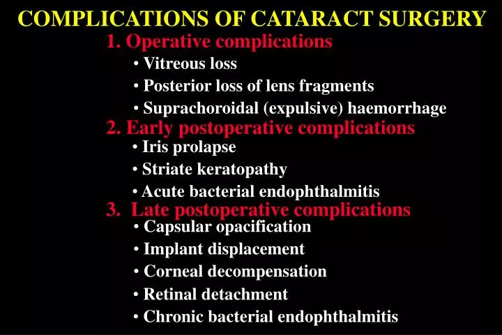
COMPLICATIONS OF CATARACT SURGERY. 1. Operative complications. Vitreous loss. Posterior loss of lens fragments. Suprachoroidal (expulsive) haemorrhage. 2. Early postoperative complications. Iris prolapse. Striate keratopathy. Acute bacterial endophthalmitis.

E N D
COMPLICATIONS OF CATARACT SURGERY 1. Operative complications • Vitreous loss • Posterior loss of lens fragments • Suprachoroidal (expulsive) haemorrhage 2. Early postoperative complications • Iris prolapse • Striate keratopathy • Acute bacterial endophthalmitis 3. Late postoperative complications • Capsular opacification • Implant displacement • Corneal decompensation • Retinal detachment • Chronic bacterial endophthalmitis
Operative complications of vitreous loss Management Sponge or automated anterior vitrectomy Insertion of PC-IOL if adequate casular support present
Insertion of AC-IOL If adequate capsular support absent 1. Constriction of pupil 4. Coating of IOL with viscoelastic substance 2. Peripheral iridectomy 3. Glide insertion 5. Insertion of IOL 6. Suturing of incision
Management of posterior loss of lens fragments Fragments consisting of 25% or more of lens should be removed Pars plana vitrectomy and removal of fragment
Management of suprachoroidal (expulsive) haemorrhage Close incision and administer hyperosmotic agent Subsequent treatment after 7-14 days • Drain blood • Pars plana vitrectomy • Air-fluid exchange
Early postoperative complications Iris prolapse Cause • Usually inadequate • suturing of incision • Most frequently follows • inappropriate management • of vitreous loss Treatment • Excise prolapsed iris tissue • Resuture incision
Striate keratopathy Corneal oedema and folds in Descemet membrane Cause • Damage to endothelium • during surgery Treatment • Most cases resolve • within a few days • Occasionally persistent • cases may require • penetrating • keratoplasty
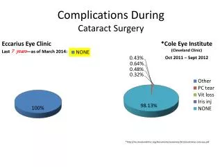
Acute bacterial endophthalmitis Incidence - about 1:1,000 • Common causative • organisms • Staph. epidermidis • Staph. aureus • Pseudomonas sp. Source of infection • Patient’s own external • bacterial flora is most • frequent culprit • Contaminated solutions • and instruments • Environmental flora including • that of surgeon and • operating room personnel
Preoperative prophylaxis Treatment of pre-existing infections Staphylococcal blepharitis Chronic conjunctivitis Chronic dacryocystitis Infected socket
Peroperative prophylaxis Meticulous prepping and draping Postoperative injection of antibiotics Instillation of povidone-iodine
Signs of severe endophthalmitis • Pain and marked visual loss • Absent or poor red reflex • Corneal haze, fibrinous exudate and • hypopyon • Inability to visualize fundus with • indirect ophthalmoscope
Signs of mild endophthalmitis • Mild pain and visual loss • Small hypopyon • Fundus visible with indirect • ophthalmoscope • Anterior chamber cells
Differential diagnosis of endophthalmitis Uveitis associated with retained lens material Sterile fibrinous reaction • No pain and few if any anterior cells • No pain or hypopyon • Posterior synechiae may develop
Management of Acute Endophthalmitis 1. Preparation of intravitreal injections 2. Identification of causative organisms • Aqueous samples • Vitreous samples 3. Intravitreal injections of antibiotics 4. Vitrectomy - only if VA is PL 5. Subsequent treatment
Preparation for sampling and injections Antibiotics Mini vitrector
Sampling and injections (1) Insert mini vitrector Make partial-thickness sclerotomy 3 mm behind limbus
Sampling and injections ( 2 ) • Insert needle attached to syringe • containing antibiotics • Remove vitrector and needle • Aspirate 0.3 ml with vitrector • Inject subconjunctival antibiotics • Give first injection of antibiotics • Disconnect syringe from needle • Give second injection
Subsequent Treatment 1. Periocular injections • Vancomycin 25 mg with ceftazidime 100 mg • or gentamicin 20 mg with cefuroxime 125 mg • Betamethasone 4 mg (1 ml) 2. Topical therapy • Fortified gentamicin 15 mg/ml and vancomycin 50 mg/ml drops • Dexamethasone 0.1% 3. Systemic therapy • Antibiotics are not beneficial • Steroids only in very severe cases
Types of capsular opacification Elschnig pearls Fibrosis • Proliferation of lens epithelium • Usually occurs within 2-6 months • May involve remnants of anterior • capsule and cause phimosis • Occurs after 3-5 years
Treatment of capsular opacification Nd:YAG laser capsulotomy • Accurate focusing is vital • Apply series of punctures • in cruciate pattern (a-c) • 3 mm opening is adequate (d) Potential complications • Damage to implant • Cystoid macular oedema • - uncommon • Retinal detachment • - rare except in high myopes
Implant displacement Decentration Optic capture • Reposition may be necessary • May occur if one haptic is inserted • into sulcus and other into bag • Remove and replace if severe
Corneal decompensation Predispositions Treatment • Penetrating keratoplasty in severe cases • Anterior chamber implant • Guarded visual prognosis because • of frequently associated CMO • Fuchs endothelial dystrophy
Retinal detachment risk factors Disruption of posterior capsule Lattice degeneration • Intraoperative vitreous loss • Treat prophylactically before or • soon after surgery • Laser capsulotomy, particularly • in high myopia
Chronic bacterial endophthalmitis Signs • Late onset, persistent, low-grade • uveitis - may be granulomatous • Low virulence organisms trapped • in capsular bag • Commonly caused by P. acnes or Staph. • epidermidis • White plaque on posterior capsule
Treatment of chronic endophthalmitis • Recurrence after cessation of treatment • Initially good response to topical • steroids • Inject intravitreal vancomycin • Remove IOL and capsular bag if • unresponsive