Download
1 / 21
270 likes | 620 Views
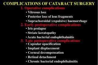
postoperative cataract complications. Lecture 13 Liana Al-Labadi, O.D. Complications. Corneal Edema. Mild Uveitis. Seidel’s Sign. Endophthalmitis. Rarely occurs after modern cataract surgery Visually threatening condition Carries very poor prognosis- 50% blindness if treatment is delayed

E N D
postoperative cataract complications • Lecture 13 • Liana Al-Labadi, O.D.
Endophthalmitis • Rarely occurs after modern cataract surgery • Visually threatening condition • Carries very poor prognosis- 50% blindness if treatment is delayed • Can present as an acute form or chronic form • Symptoms: • Mild to severe pain • Redness • Loss of vision • Floaters • Photophobia • Signs: • The hallmark of endophthalmitis is vitreous inflammation • Eyelid & periorbital edema • Chemosis • Corneal edema • AC reaction • Hypopyon • Etiology: • Toxic material introduced to the eye • Poor sterlization- materials, injection needle, surgeon, nurses • Management: • Culture- must identify organism type • IV AB & hospitalization
Pseudophakic Bullous Keratopathy (PBK) • PBK is a post-op condition that can occur as a complication following PE PCIOL • May present early or it may not present for many years • Symptoms: • Decreased vision • Pain • FBS & tearing • Photophobia • red eye • Signs: • Mild to marked persistent stromal edema in an eye in which the native lens has been removed • Increased K thickness • Bullous formation in severe cases--> these rupture & cause pain • Descemet folds • K vascularization • CME may be present
Pseudophakic Bullous Keratopathy (PBK) • Etiology: Due to compromised endothelial cell function • Both intraoperative insult to the endothelium and long-term cell damage as a result of the lens implant can lead to PBK • Proposed mechanism to endothelial cell loss include: • Direct trauma during surgery • Prolonged irrigation • Toxic medications • Persistent inflammation • Increased IOP • AC IOL- associated with more endothelial cell loss than PC IOL • “Intermittent touch” between IOL & K • Chronic low-grade inflammation caused by IOL haptics or footplates • May disrupt the normal flow of aqueous in the AC which affects the nutrient flow to endothelial cells • Management: • Mild PBK • Hypertonic saline drops (Muro 128-5% sodium chloride) • Steroids • BCL • PK or DSAEK • Prevention: • Use of preoperative endothelial cell counts in high risk cases • Use of viscoelastic solution during the surgery
PBK http://emedicine.medscape.com/article/1193218-overview http://www.doctorshangout.com/photo/bullous-keratopathy http://flylib.com/books/en/3.283.1.8/1
Posterior Capsular opacification (PCO) • After ECCE & PE, proliferation of lens epithelial cells can lead to posterior capsule opacification • Posterior capsule becomes opacified as a result of continued proliferation of lens cells from the residual anterior lens epithelium or from residual fibrosis that could not be removed at the time of surgery • Occurs in 50% of patients within 5 years after ECCE surgery • Occurs in 1/5 people who undergo PE PCIOL • Symptoms: decreased vision & FBS & pain if bullae present • Signs: • Blurry vision • Glare “secondary cataracts” • Asymptomatic • Management: • YAG laser capsulotomy • Done when the patient is symptomatic • Follow up in 1 week then 1 month s/p YAG • Complications: • Increased IOP • Damage to IOL • IOL dislocation • Inflammatory reaction • CME • RD- especially in myopic patients (1-3% of patients)
PCO http://flylib.com/books/en/3.283.1.8/1/ http://flylib.com/books/en/3.283.1.8/1/
PCO http://dro.hs.columbia.edu/pco2.htm
Cystoid macular edema (CME) • A condition in which fluid accumulates within the sensory retina in the macular area • May occur after intraocular surgery • Cataract • Filtration procedures • RD surgery • Associated with other systemic & ocular conditions including: • Diabetes • Peripheral uveitis • RP • May occur in as high as 20% of cataract surgeries, but only persists in 1-2% • Onset: 6-10 wks s/p CE • Symptoms: decreased hazy vision • Signs: • Hyperopic shift in RE • Macular haze • Petaloid appearance on FA is the hallmark of CME (or flower petal) • Evidence suggests inflammation plays a role • Management: • May improve without treatment if no other surgical complications • 70% of post-CE CME resolves spontaneously within 6 months • NSAIDs • CME may be recurrent • CME secondary to CE is AKA Irvin-Grass Syndrome
CME http://dro.hs.columbia.edu/pco2.htm http://dro.hs.columbia.edu/pco2.htm http://www.retinatexas.com/images/CSME.jpg
CME http://flylib.com/books/en/3.283.1.8/1/
Bye Bye Cataract- conclusion • Approach cases in a conservative manner • Correlate VA with anterior segment appearance • Maintain communication with surgeon and family physicians • Make referrals to experienced modern surgeons