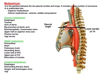
Download
1 / 8
90 likes | 397 Views
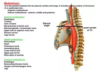
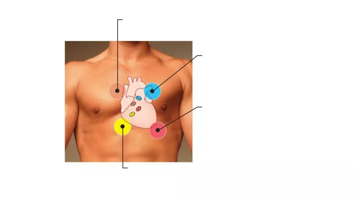
Aortic valve sounds heard in 2nd intercostal space at right sternal margin. Pulmonary valve sounds heard in 2nd intercostal space at left sternal margin. Mitral valve sounds heard over heart apex (in 5th intercostal space) in line with middle of clavicle.

E N D
Aortic valvesounds heard in 2nd intercostal space at right sternal margin Pulmonary valve sounds heard in 2nd intercostal space at left sternal margin Mitral valvesounds heard over heart apex (in 5th intercostal space) in line with middle of clavicle Tricuspid valvesounds typically heard in right sternal margin of 5th intercostal space
Anatomy of the intrinsic conduction system showing the sequence of electrical excitation
Thesinoatrial (SA) node(pacemaker) 1 The impulses pause (0.1 s) at the atrioventricular (AV) node. 2 ThePurkinje fibers Theatrioventricular (AV) bundle 3 Thebundle branches 4 ThePurkinje fibers 5 Anatomy of the intrinsic conduction system showing the sequence of electrical excitation
QRS complex Sinoatrial node Atrioventricular node S-T Segment P-Q Interval Q-T Interval
QRS complex Sinoatrial node Ventricular depolarization Ventricular repolarization Atrial depolarization Atrioventricular node S-T Segment P-Q Interval Q-T Interval
Left heart “Lup” = “Dup” = Electrocardiogram Heart sounds Pressure (mm Hg) EDV = ESV = SV = Ventricular volume (ml) Atrial _________ Atrioventricular valves Open Closed Open Aortic and pulmonary valves Closed Open Closed Ventricular _________ Ventricular _________
Left heart QRS “Lup” = closing of L AV valve or Mitral valve (1st heart sound) “Dup” = closing of Aortic valve (2nd heart sound) P T P Electrocardiogram 1st 2nd Heart sounds Dicrotic notch Aorta Pressure (mm Hg) Left ventricle Atrial systole Left atrium EDV = End Diastolic Volume ESV = End Systolic Volume SV = Stroke Volume EDV Ventricular volume (ml) SV Atrial SYSTOLE ESV Atrioventricular valves Open Closed Open Aortic and pulmonary valves Closed Open Closed Ventricular SYSTOLE Ventricular DIASTOLE