Download
1 / 51
670 likes | 1.84k Views
NEUROMUSCULAR JUNCTION. Dr. Sidra Hamid Physiology Department. CASE 4: 35 year old woman with progressive muscle weakness.

E N D
NEUROMUSCULAR JUNCTION Dr. Sidra Hamid Physiology Department
CASE 4: 35 year old woman with progressive muscle weakness • A 35 years old woman resident of Rawalpindi presented in foundation OPD with progressive weakness for the last 2 months. She has also noticed intermittent drooping of both of her eye lids, and progressive facial muscles weakness while speaking. She also complaints of weakness and tiredness while climbing the stairs of her office has difficulty while typing a lengthy official replies to their clients.
Her general physical examination revealed a pulse of 82/min. B.P 120/80 mm of Hg. Temp. 98 F and Resp. rate 16/min. with drooping of both eyelids ( Ptosis +ive). Her laboratory investigations revealed positive anti-choline receptor antibodies. Rest of laboratory workup was unremarkable.
LEARNING OBJECTIVES • Describe the physiological anatomy of Neuromuscular Junction (NMJ). • Terminal button. • Motor end plate. • Motor End Plate potential and how action potential is generated in muscle. • Synaptic trough/ gutter/ cleft. • Chemicals/ drugs/ diseases effecting neuromuscular transmission
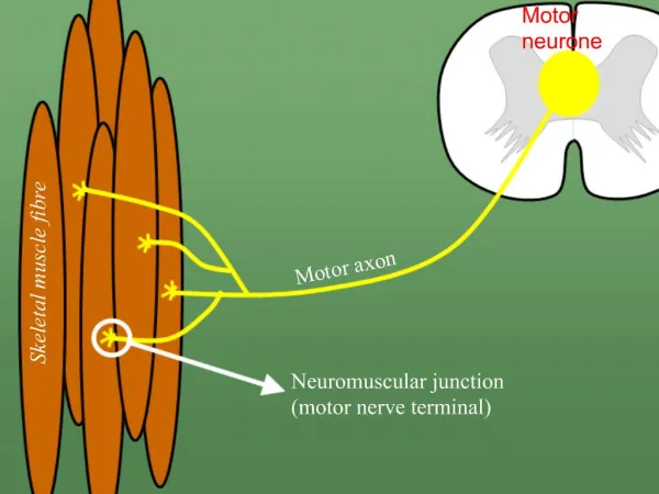
DEFINITION “ The place where the motor neuron makes a functional contact with the skeletal muscle cell is called NEUROMUSCULAR JUNCTION or MYONEURAL JUNCTION”
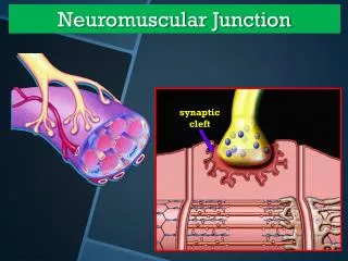
Neuromuscular Junction • Neuromuscular Junction A neuromuscular junction exists between a motor neuron and a skeletal muscle. - Synapse A junction between two neurons
INNERVATION OF SKELETAL MUSCLE FIBERS • Large, myelinated nerve fibers • Originate from large motor neurons in the anterior horns of the spinal cord • Each nerve fiber, branches and stimulates from three to several hundred skeletal muscle fibers • The action potential initiated in the muscle fiber by the nerve signal travels in both directions toward the muscle fiber ends
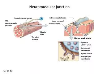
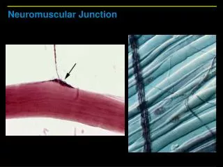
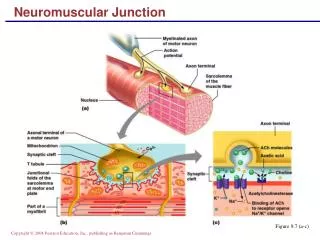
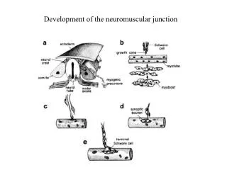
MOTOR END PLATE • The nerve fiber forms a complex of branching nerve terminals that invaginate into the surface of the muscle fiber but lie outside the muscle fiber plasma membrane • Entire structure - motor endplate. • Covered by one or more Schwann cells that insulate it from the surrounding fluids.
AXON TERMINAL • SYNAPTIC VESICLES • Size 40 nanometers • Formed by the Golgi apparatus in the cell body of the motor neuron in the spinal cord. • Transported by axoplasm to the neuromuscular junction at the tips of the peripheral nerve fibers. • About 300,000 of these small vesicles collect in the nerve terminals of a single skeletal muscle end plate.
MITOCHONDRIA • Numerous • Supply ATP • Energy source for synthesis of excitatory neurotransmitter, acetylcholine • DENSE BARS • Present on the inside surface of neural membrane
VOL TAGE GATED CALCIUM CHANNELS • Protein particles that penetrate the neural membrane on each side 0f dense bar • When an action potential spreads over the terminal, these channels open and calcium ions diffuse to the interior of the nerve terminal. • The calcium ions, exert an attractive influence on the acetylcholine vesicles, drawing them to the neural membrane adjacent to the dense bars.
The vesicles then fuse with the neural membrane and empty their acetylcholine into the synaptic space by the process of exocytosis • Calcium acts as an effective stimulus for causing acetylcholine release from the vesicles • Acetylcholine is then emptied through the neural membrane adjacent to the dense bars and binds with acetylcholine receptors in the muscle fiber membrane
MUSCLE FIBER MEMBRANE • SYNAPTIC TROUGH • The muscle fiber membrane where it is invaginated by a nerve terminal and a depression is formed • SYNAPTIC CLEFT • The space between the nerve terminal and the fiber membrane is called the synaptic space or synaptic cleft
SUBNEURAL CLEFT • Numerous smaller folds of the muscle membraneat the bottom of the gutter • Greatly increase the surface area. • ACETYLCHOLINE RECEPTORS • Acetylcholine-gated ion channels • Located almost entirely near the mouths of the sub neural clefts lying immediately below the dense bar areas
ACETYLCHOLINE RECEPTORS • Acetylcholine-gated ion channels • Molecular weight -275,000
SUBUNITS • Two alpha, one each of beta, delta, and gamma • Penetrate all the way through the membrane • Lie side by side in a circle- form a tubular channel • Two acetylcholine molecules attach to the two alpha subunits, opens the channel • RESTING STATE • 2 Ach molecules not attached to the alpha subunit • Channel remains constricted
OPENED Ach CHANNEL • 2 Ach molecules attached to the alpha subunit of receptor • Diameter- 0.65 nanometer • Allows important positive ions—SODIUM, potassium, and calcium to move easily through the opening. • Disallows negative ions, such as chloride to pass through because of strong negative charges in the mouth of the channel that repel these negative ions.
SODIUM IONS • Far more sodium ions flow through the acetylcholine channels to the inside than any other ions • The very negative potential on the inside of the muscle membrane, –80 to –90 mili volts, pulls the positively charged sodium ions to the inside of the fiber • Simultaneously prevents efflux of the positively charged potassium ions when they attempt to pass outward
END PLATE POTENTIAL • Opening the acetylcholine-gated channels allows large numbers of sodium ions to pour to the inside of the fiber • Sodium ions carry with them large numbers of positive charges • Creates a local positive potential change inside the muscle fiber membrane, called the end plate potential. • End plate potential initiates an action potential that spreads along the muscle membrane • Causes muscle contraction
Events of Neuromuscular Junction • Propagation of an action potential to a terminal button of motor neuron. • Opening of voltage-gated Ca2+ channels. • Entry of Calcium into the terminal button. • Release of acetylcholine (by exocytosis). • Diffusion of Ach across the space. • Binding of Ach to a receptor on motor end plate.
Examples of Chemical Agents and Diseases that Affect the Neuromuscular Junction Mechanism Chemical Agent or Disease Alters Release of Acetylcholine * Cases explosive release of acetylcholine * Black widow spider venom * Blocks release of acetylcholine * Clostridium botulinum toxin Block acetylcholine Receptor * Bind reversibly * Curare * Auto antibodies inactivate acetylcholine * Myasthenia gravis receptors Prevents inactivation of acetylcholine * Irreversibly inhibits acetylcholinesterase * Organophosphates * Temporary inhibits acetylcholinesterase * Neostigmine