Neuromuscular Junction
500 likes | 1.27k Views
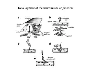
b. Neuromuscular Junction. Conscious thought (to move a muscle) results in activation of a motor neuron, and release of the neurotransmitter acetylcholine (AcCh) at the NM junction The enzyme acetylcholinesterase breaks down AcCh after a short period of time.

Neuromuscular Junction
E N D
Presentation Transcript
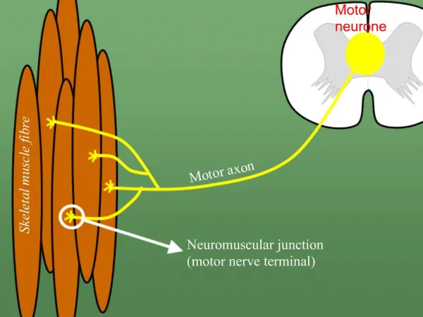
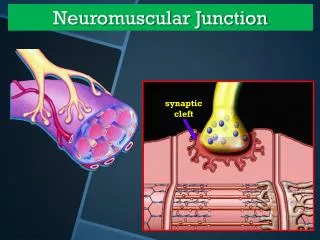
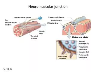
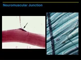
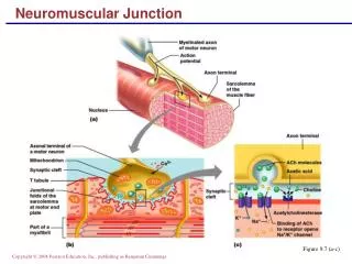
Neuromuscular Junction • Conscious thought (to move a muscle) results in activation of a motor neuron, and release of the neurotransmitter acetylcholine (AcCh) at the NM junction • The enzyme acetylcholinesterase breaks down AcCh after a short period of time
Neuromuscular Junction • The plasma membrane on the “far side” of the NMJ belongs to the muscle cell and is called the motor end plate • The motor end plate is rich in chemical (ligand) - gated sodium channels that respond to AcCh. Another way to say this: The receptors for AcCh are on the ligand-gated sodium channels on the motor end plate
Neuromuscular Junction • The chemical events at the NMJ transmit the electrical events of a neuronal action potential into the electrical events of a muscle action potential
Neuromuscular JunctionInteractions Animation • Neuromuscular Junctions You must be connected to the internet to run this animation.
Muscle Action Potential • The muscle AP is propagated over the surface of the muscle cell membrane (sarcolemma) via voltage (electrical)-gated Na+ and K+ channels
Muscle Action Potential • By placing a micropipette inside a muscle cell, and then measuring the electrical potential across the cell membrane, the phases of an action potential (AP) can be graphed (as in this figure)
Muscle Action Potential • The behavior of the Na+ and K+ channels, at various points in the AP, are seen in this graphic • Na+ gates open during the depolarization phase • K+ gates open during the repolarization phase
Generating An Action Potential • The flow of ions through cell a membrane looks a lot like a "piece" of electricity flowing through a wire (but not as fast) • Generating an AP on the muscle membrane involves the transfer of information from an electrical signal (down the neuron), to a chemicalsignal (at the NMJ), back to an electrical signal (depolarization of the sarcolemma) • This added complexity (changing from electrical to chemical back to electrical signals) provides necessary control of the process
Excitation-Contraction Coupling • EC coupling involves putting it all together • The thought process going on in the brain • The AP arriving at the neuromuscular junction • The regeneration of an AP on the muscle membrane • Release of Ca2+ from the sarcoplasmic reticulum • Sliding of thick on thin filaments in sarcomeres • Generation of muscle tension (work)
Excitation-Contraction Coupling • Role Players in E-C coupling • Regenerate AP • The T-tubules • The SR • Ca2+ release • Troponin/Tropomyosin • ATP • Myosin binding • Filaments slide • Muscles contract • The brain • The motor neuron • Acetylcholine (ACh) • Acetylcholinesterase enzyme • Ach receptors on the • motor endplate • Na+-K+ channels on the sarcolemma • Na+ flow • K+ flow
Contraction of SarcomereInteractions Animation • Contraction of a Sarcomere You must be connected to the internet to run this animation.
Sources of Muscle Energy • Stored ATP • 3 seconds • Energy transferred from stored creatine phosphate • 12 seconds • Aerobic ATP production • Anaerobic glucose use • 30-40 seconds
Skeletal Muscle Metabolism • In a state of homeostasis, muscle use of O2 and nutrients is balanced by the production of manageable levels of waste products like • CO2 • Heat - 70-80% of the energy used by muscles is lost as heat - muscle activity is important for maintaining body temperature • Lactic acid (anaerobic)
Skeletal Muscle Metabolism • Oxygen Debt, or "Excess Post-Exercise Oxygen Consumption" (EPOC) is the amount of O2 repayment required after exercise in skeletal muscle to: • Replenish ATP stores • Replenish creatine phosphate and myoglobin stores • Convert lactic acid back into pyruvate so it can be used in the Krebs cycle to replenish ATP
Muscle Metabolism • Muscle Metabolism You must be connected to the internet to run this animation.
Cardiac and Smooth Muscle Metabolism • In response to a single AP, cardiac muscle contracts 10-15 times longer than skeletal muscle, and must continue to do so, without rest, for the life of the individual • To meet this constant demand, cardiac muscle generally uses the rich supply of O2 delivered by the extensive coronary circulation to generate ATP through aerobic respiration
Cardiac and Smooth Muscle Metabolism • Like cardiac muscle, smooth muscle (in your deep organs) is autorhythmic and is not under voluntary control (your heart beats and your stomach digests without you thinking about it). • Unlike cardiac (and skeletal muscle) however, smooth muscle has a low capacity for generating ATP and does so only through anaerobic respiration (glycolysis)
The Motor Unit • Motor Unit is composed of a motor neuron plus all of the muscle cells it innervates • High precision • Fewer muscle fibers per neuron • Laryngeal and extraocular muscles (2-20) • Low precision • Many muscle fibers per neuron • Thigh muscles (2,000-3,000)
The Motor Unit Florescent dye is used to show the terminal processes of a single neuron which terminate on a few muscle fibers
The Motor Unit Activities requiring extreme precision (like the subtle and rapid movements of the eye) involve muscles with very small motor units (1-4 muscle fibers/neuron)
The Motor Unit • All-or-none principle of muscle contraction • When an individual muscle fiber is stimulated to depolarization, and an action potential is propagated along its sarcolemma, it must contract to it’s full force—it can’t partially contract • Also, when a single motor unit is recruited to contract, all the muscle fibers in that motor unit must all contract at the same time
Skeletal Muscle Fiber Types • Skeletal muscle fibers are not all alike in appearance or function. By appearance: • Red muscle fibers (the dark meat in chicken legs) have a high myoglobin content, more mitochondria, more energy stores, and a greater blood supply • White muscle fibers (the white meat in chicken breasts) have less myoglobin, mitochondria, and blood supply
Skeletal Muscle Fiber Types • Slow oxidative fibers (SO) are small, appear dark red, are the least powerful type. They are very fatigue resistant • Used for endurance like running a marathon • Fast oxidative-glycolytic fibers (FOG) are intermediate in size, appear dark red, and are moderately resistant to fatigue. Used for walking • Fast glycolytic fibers (FG)are large, white, and powerful • Suited to intense anaerobic activity of short duration
Skeletal Muscle Fiber Types • Most skeletal muscles are a mixture of all three types of skeletal muscle fibers; about half the fibers in a typical skeletal muscle are slow oxidative (SO) fibers • Within a particular motor unit all the skeletal muscle fibers are the same type • The different motor units in a muscle are recruited in a specific order depending on the task being performed (fast anaerobic activity for maximal force, etc.)
Tension in a Muscle • There is a brief delay called the latent period as the AP sweeps over the sarcolemma and Ca2+ions are released from the sarcoplasmic reticulum (SR) • During the next phase the fiber is actively contracting • This is followed by relaxation as the Ca2+ions are re-sequestered into the SR and myosin binding sites are covered by tropomyosin • Temporary loss of excitability is call the refractory period – All muscle fibers in a motor unit will not respond to a stimulus during this short time
Tension in a Muscle • A twitch is recorded when a stimulus that results in contraction (force) of a single muscle fiber is measured over a very brief millisecond time frame
Tension in a Muscle • Applying increased numbers of action potentials to a muscle fiber (or a fascicle, a muscle, or a muscle group) results in fusion of contractions (tetanus) and the performance of useful work
Tension in a Muscle • Two motor units, one in green, the other in purple, demonstrate the concept of progressive activation of a muscle known as recruitment • Recruitmentallows a muscle to accomplish increasing gradations of contractile strength
Muscle TensionInteractions Animation • Control of Muscle Tension You must be connected to the internet to run this animation.
Muscle Contraction • Isotonic contractions results in movement • Concentric isotonic is a type of muscle contraction in which the muscle shorten while generating force • Eccentric isotonic is a contraction in which muscle tension is less than the resistance (the muscle lengthens) • Isometric contractions results in no movement • Muscle force and resistance are equal • Supporting objects in a fixed position and posture
Imbalances of Homeostasis • Exercise-induced muscle damage • After intense exercise electron micrographs reveal considerable muscle damage including torn sarcolemmas and disrupted Z-discs • Blood levels of proteins normally confined only to muscle (including myoglobin and the enzyme creatine kinase) increase as they are released from damaged muscle
Imbalances of Homeostasis • Spasm • A sudden involuntary contraction of a single muscle within a large group of muscles – usually painless • Cramp • Involuntary and often painful muscle contractions • Caused by inadequate blood flow to muscles (such as in dehydration), overuse and injury, and abnormal blood electrolyte levels
Imbalances of Homeostasis • Disease States and Disorders • Fibrosis (myofibrosis) • Replacement of muscle fibers by excessive amounts of connective tissues (fibrous scar tissue) • Myosclerosis • Hardening of the muscle caused by calcification • Both myosclerosis and muscle fibrosis occur as a result of trauma and various metabolic disorders
Imbalances of Homeostasis • Aging • In part due to decreased levels of physical activity, with aging humans undergo a slow, progressive loss of skeletal muscle mass that is replaced largely by fibrous connective tissue and adipose tissue • Muscle strength at 85 is about half that at age 25 • Compared to the other two fiber types, the relative number of slow oxidative fibers appears to increase