Download
1 / 59
590 likes | 592 Views
Learn about the 5 elements of a reflex arc, the difference between a reflex and a voluntary reaction, and the major regions of the human brain including the cerebral hemispheres.

E N D
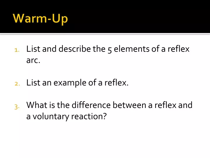
Warm-Up • List and describe the 5 elements of a reflex arc. • List an example of a reflex. • What is the difference between a reflex and a voluntary reaction?
4 Major Regions • Cerebral Hemispheres • Diencephalon • Brain stem • Cerebellum
1. Cerebral Hemispheres (Cerebrum) • L & R hemispheres • Corpus callosum: large fiber tract; connects 2 hemispheres • Lobes: major regions (named for cranial bones) • Parietal, frontal, occipital, temporal • Gyri (gyrus) = elevated ridges of tissue • Sulci (sulcus) = shallow grooves • Fissures = deeper grooves, separate large regions of brain • Motor & sensory function: opposite hemispheres
Cerebral Cortex • Grey matter • “Executive suite” conscious mind
Cerebral Hemispheres • Surface markings • Central sulcus • Separates the precentral gyrus of the frontal lobe and the postcentral gyrus of the parietal lobe • Longitudinal fissure • Separates the two hemispheres • Transverse cerebral fissure • Separates the cerebrum and the cerebellum
Precentral gyrus Central sulcus Postcentral gyrus Frontal lobe Parietal lobe Parieto-occipital sulcus (on medial surface of hemisphere) Lateral sulcus Occipital lobe Temporal lobe Transverse cerebral fissure Cerebellum Pons Medulla oblongata Fissure Spinal cord (a deep sulcus) Gyrus Cortex (gray matter) Sulcus White matter (a) Figure 12.6a
Cerebral Cortex • Thin (2–4 mm) superficial layer of gray matter • 40% of the mass of the brain • Site of conscious mind: awareness, sensory perception, voluntary motor initiation, communication, memory storage, understanding
Functional Areas of the Cerebral Cortex • The three types of functional areas are: • Motor areas—control voluntary movement • Sensory areas—conscious awareness of sensation • Association areas—integrate diverse information • Conscious behavior involves the entire cortex
Motor Areas • Primary (somatic) motor cortex • Premotor cortex • Broca’s area • Frontal eye field
Motor areas Sensory areas and related association areas Central sulcus Primary motor cortex Primary somatosensory cortex Premotor cortex Somatic sensation Frontal eye field Somatosensory association cortex Broca’s area (outlined by dashes) Gustatory cortex (in insula) Taste Prefrontal cortex Wernicke’s area (outlined by dashes) Working memory for spatial tasks Executive area for task management Primary visual cortex Working memory for object-recall tasks Vision Visual association area Solving complex, multitask problems Auditory association area Hearing Primary auditory cortex (a) Lateral view, left cerebral hemisphere Motor association cortex Primary sensory cortex Primary motor cortex Sensory association cortex Multimodal association cortex Figure 12.8a
Posterior Motor Anterior Motor map in precentral gyrus Toes Jaw Primary motor cortex (precentral gyrus) Tongue Swallowing Figure 12.9
Primary somatosensory cortex Somatosensory association cortex Visual areas Auditory areas Olfactory cortex Gustatory cortex Visceral sensory area Vestibular cortex Sensory Areas
Motor areas Sensory areas and related association areas Central sulcus Primary motor cortex Primary somatosensory cortex Premotor cortex Somatic sensation Frontal eye field Somatosensory association cortex Broca’s area (outlined by dashes) Gustatory cortex (in insula) Taste Prefrontal cortex Wernicke’s area (outlined by dashes) Working memory for spatial tasks Executive area for task management Primary visual cortex Working memory for object-recall tasks Vision Visual association area Solving complex, multitask problems Auditory association area Hearing Primary auditory cortex (a) Lateral view, left cerebral hemisphere Motor association cortex Primary sensory cortex Primary motor cortex Sensory association cortex Multimodal association cortex Figure 12.8a
Posterior Sensory Anterior Sensory map in postcentral gyrus Genitals Primary somato- sensory cortex (postcentral gyrus) Intra- abdominal Figure 12.9
Visual Areas • Primary visual (striate) cortex • Extreme posterior tip of the occipital lobe • Receives visual information from the retinas
Multimodal Association Areas • Receive inputs from multiple sensory areas • Send outputs to multiple areas • Allow us to give meaning to information received, store it as memory, compare it to previous experience, and decide on action to take
Multimodal Association Areas • Three parts • Anterior association area (prefrontal cortex) • Posterior association area • Limbic association area
Anterior Association Area (Prefrontal Cortex) • Most complicated cortical region • Involved with intellect, cognition, recall, and personality • Contains working memory needed for judgment, reasoning, persistence, and conscience • Development depends on feedback from social environment
Posterior Association Area • Large region in temporal, parietal, and occipital lobes • Plays a role in recognizing patterns and faces and localizing us in space • Involved in understanding written and spoken language (Wernicke’s area)
2. Diencephalon (interbrain) 3 main structures: • Thalamus: relay station for incoming info • Hypothalamus: • Autonomic control center (heart rate, BP, digestion) • Emotional response (limbic system) • Body temperature regulation • Regulate food intake • Sleep-wake cycles • Control endocrine system pituitary gland at base • Epithalamus: pineal gland (sleep-wake cycle)
Thalamus • 80% of diencephalon • Contains several nuclei, named for their location • Nuclei project and receive fibers from the cerebral cortex
Dorsal nuclei Medial Lateral dorsal Lateral posterior Pulvinar Anterior nuclear group Medial geniculate body Reticular nucleus Lateral geniculate body Ventral postero- lateral Ventral anterior Ventral lateral Ventral nuclei (a) The main thalamic nuclei. (The reticular nuclei that “cap” thethalamus laterally are depicted as curving translucent structures.) Figure 12.13a
Thalamic Function • Gateway to the cerebral cortex • Sorts, edits, and relays information • Afferent impulses from all senses and all parts of the body • Impulses from the hypothalamus for regulation of emotion and visceral function • Impulses from the cerebellum and basal nuclei to help direct the motor cortices • Mediates sensation, motor activities, cortical arousal, learning, and memory
Hypothalamus • Contains many nuclei • Example: mammillary bodies • Paired anterior nuclei • Olfactory relay stations • Infundibulum—stalk that connects to the pituitary gland
Paraventricular nucleus Dorsomedial nucleus Anterior commissure Fornix Preoptic nucleus Posterior hypothalamic nucleus Anterior hypothalamic nucleus Lateral hypothalamic area Supraoptic nucleus Ventromedial nucleus Supra- chiasmatic nucleus Mammillary body Arcuate nucleus Optic chiasma Pituitary gland Infundibulum (stalk of the pituitary gland) (b) The main hypothalamic nuclei. Figure 12.13b
Hypothalamic Function • Autonomic control center for many visceral functions (e.g., blood pressure, rate and force of heartbeat, digestive tract motility) • Center for emotional response: Involved in perception of pleasure, fear, and rage and in biological rhythms and drives
Hypothalamic Function • Regulates body temperature, food intake, water balance, and thirst • Regulates sleep and the sleep cycle • Controls release of hormones by the anterior pituitary • Produces posterior pituitary hormones
Epithalamus • Pineal gland—extends from the posterior border and secretes melatonin • Melatonin—helps regulate sleep-wake cycles
3. Brain Stem • Programmed, automatic behaviors for survival • 3 regions: • Midbrain: vision, hearing, reflex • Pons: breathing • Medulla oblongata: heart rate, BP, breathing, swallowing, vomiting, coughing, sneezing
Brain Stem • Three regions • Midbrain • Pons • Medulla oblongata
Brain Stem • Controls automatic behaviors necessary for survival • Associated with 10 of the 12 pairs of cranial nerves
Frontal lobe Olfactory bulb (synapse point of cranial nerve I) Optic chiasma Optic nerve (II) Optic tract Mammillary body Midbrain Pons Temporal lobe Medulla oblongata Cerebellum Spinal cord Figure 12.14
View (a) Optic chiasma Optic nerve (II) Diencephalon Crus cerebri of cerebral peduncles (midbrain) • Thalamus • Hypothalamus Thalamus Diencephalon Mammillary body Hypothalamus Midbrain Oculomotor nerve (III) Pons Brainstem Trochlear nerve (IV) Medulla oblongata Trigeminal nerve (V) Pons Middle cerebellar peduncle Facial nerve (VII) Abducens nerve (VI) Vestibulocochlear nerve (VIII) Glossopharyngeal nerve (IX) Hypoglossal nerve (XII) Pyramid Vagus nerve (X) Ventral root of first cervical nerve Accessory nerve (XI) Decussation of pyramids Spinal cord (a) Ventral view Figure 12.15a
Crus cerebri of cerebral peduncles (midbrain) Thalamus View (b) Infundibulum Superior colliculus Pituitary gland Inferior colliculus Trochlear nerve (IV) Superior cerebellar peduncle Trigeminal nerve (V) Pons Middle cerebellar peduncle Facial nerve (VII) Inferior cerebellar peduncle Abducens nerve (VI) Vestibulocochlear nerve (VIII) Glossopharyngeal nerve (IX) Olive Hypoglossal nerve (XII) Thalamus Vagus nerve (X) Diencephalon Hypothalamus Midbrain Accessory nerve (XI) Pons Brainstem Medulla oblongata (b) Left lateral view Figure 12.15b
Pons • Connect higher brain centers and the spinal cord • Relay impulses between the motor cortex and the cerebellum • Origin of cranial nerves V (trigeminal), VI (abducens), and VII (facial) • Nuclei that help maintain normal rhythm of breathing
Medulla Oblongata • Joins spinal cord at foramen magnum • Relay sensory information from muscles and joints to cerebellum • Cranial nerves VIII, X, and XII are associated with the medulla • Mediates responses that maintain equilibrium
Medulla Oblongata • Autonomic reflex centers • Cardiovascular center • Cardiac center adjusts force and rate of heart contraction • Vasomotor center adjusts blood vessel diameter for blood pressure regulation • Respiratory centers • Generate respiratory rhythm • Control rate and depth of breathing
Medulla Oblongata • Additional centers regulate • Vomiting • Hiccuping • Swallowing • Coughing • Sneezing
Fourth ventricle Solitary nucleus Choroid plexus Hypoglossal nucleus (XII) Dorsal motor nucleus of vagus (X) Vestibular nuclear complex (VIII) Inferior cerebellar peduncle Cochlear nuclei (VIII) Lateral nuclear group Nucleus ambiguus Medial nuclear group Reticular formation Inferior olivary nucleus Raphe nucleus Pyramid Medial lemniscus (c) Medulla oblongata Figure 12.16c
4. Cerebellum • Balance, equilibrium, timing of skeletal muscle activity