Download
1 / 1
10 likes | 72 Views
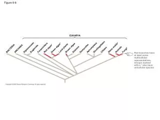
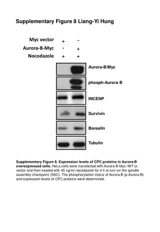
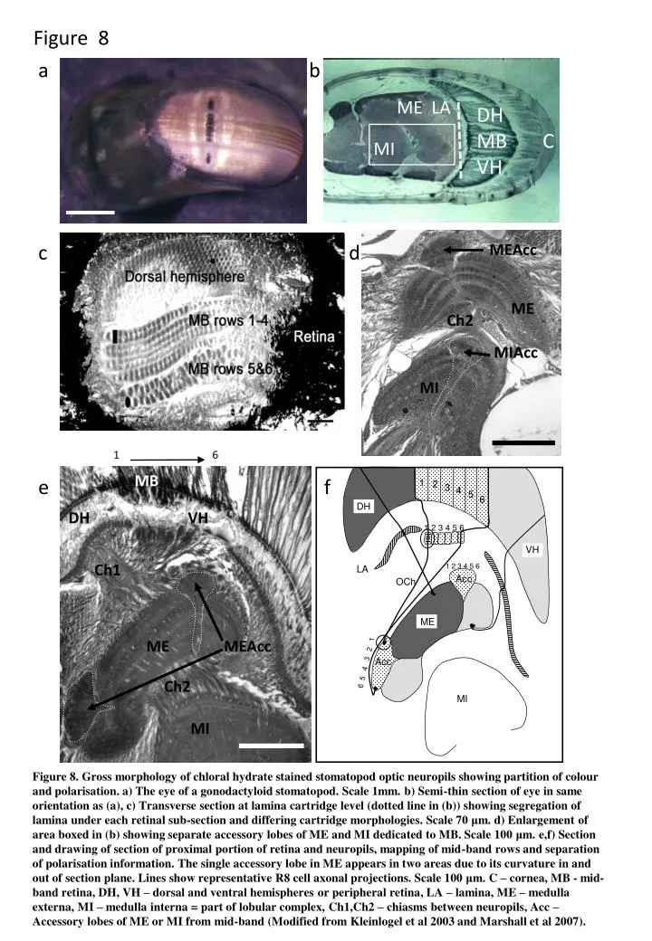
1. 6. MB. DH VH. Ch1. ME. MEAcc. Ch2. MI. Figure 8. a b c d e f. 1. 2. 3. 4. ME LA. 5. DH MB VH. 6. DH. C. 1 2 3 4 5 6. MI. VH. 1 2 3 4 5 6. LA. Acc. OCh. ME. MEAcc. Acc. 6 5 4 3 2 1. ME. MI. Ch2.

E N D
1 6 MB DH VH Ch1 ME MEAcc Ch2 MI Figure 8 a b c d e f 1 2 3 4 ME LA 5 DH MB VH 6 DH C 1 2 3 4 5 6 MI VH 1 2 3 4 5 6 LA Acc OCh ME MEAcc Acc 6 5 4 3 2 1 ME MI Ch2 MIAcc MIAcc MI Figure 8. Gross morphology of chloral hydrate stained stomatopod optic neuropils showing partition of colour and polarisation. a) The eye of a gonodactyloid stomatopod. Scale 1mm. b) Semi-thin section of eye in same orientation as (a), c) Transverse section at lamina cartridge level (dotted line in (b)) showing segregation of lamina under each retinal sub-section and differing cartridge morphologies. Scale 70 μm. d) Enlargement of area boxed in (b) showing separate accessory lobes of ME and MI dedicated to MB. Scale 100 μm. e,f) Section and drawing of section of proximal portion of retina and neuropils, mapping of mid-band rows and separation of polarisation information. The single accessory lobe in ME appears in two areas due to its curvature in and out of section plane. Lines show representative R8 cell axonal projections. Scale 100 μm. C – cornea, MB - mid-band retina, DH, VH – dorsal and ventral hemispheres or peripheral retina, LA – lamina, ME – medulla externa, MI – medulla interna = part of lobular complex, Ch1,Ch2 – chiasms between neuropils, Acc – Accessory lobes of ME or MI from mid-band (Modified from Kleinlogel et al 2003 and Marshall et al 2007).