Download
1 / 59
630 likes | 883 Views
Distinct memory systems mediating declarative, emotional and procedural memory functions. AIO Course – Cognitive Neurosciences January 28, 2002. Distinct memory systems mediating declarative, emotional and procedural memory functions.

E N D
Distinct memory systems mediating declarative, emotional and proceduralmemory functions AIO Course – Cognitive Neurosciences January 28, 2002
Distinct memory systems mediating declarative, emotional and proceduralmemory functions In this lecture the general features of the multiplicity of memory systems that subserve distinct categories of memory functions are described. The experimental paradigms that are used to study declarative and nondeclarative (procedural) learning are analyzed with especial emphasis on animal models of associative learning, i.e. classical and emotional conditioning and of declarative memory. The corresponding anatomic circuits and neuronal elements are considered in general terms. The neurobiological mechanisms that likely underlie the formation of memory traces in such neuronal circuits at the cellular and molecular levels (long-term synaptic potentiation and depression) are examined. How these mechanisms relate to the processes of encoding and retrieval of information in the brain are critically discussed.
1st cell is inscribed: “Sensus communis, imaginativa, fantasia”; 2nd cell: “cogitatia, estimatia”; 3rd cell: “memorativa”. Brain cell diagram of Gregor Reich (1503)
Distinct memory systems mediating declarative, emotional and proceduralmemory functions • Main anatomic memory systems: • Declarative memory; • Emotional memory; fear conditioning; • Motor conditioning; • Skills and Priming.
Distinct memory systems mediating declarative, emotional and proceduralmemory functions • Main anatomic memory systems: • Declarative memory; • Emotional memory; fear conditioning; • Motor conditioning; • Skills and Priming.
MTL: (para)hippocampal cortex Taxonomy of Memory Declarative (explicit) Procedural (implicit) Episodic (working) Semantic (reference) Skills Priming Conditioning Non-associative Emotional responses Motor responses MTL: (para)hippocampal cortex Reflex paths Striatum Cortex Amygdala Cerebellum & Diencephalon (Adapted from L.R. Squire: “Memory and the Brain”, 1987)
MTL: (para)hippocampal cortex Taxonomy of memory Declarative (explicit) Procedural (implicit) Episodic (working) Semantic (reference) Skills Priming Conditioning Non-associative Emotional responses Motor responses MTL: (para)hippocampal cortex Reflex paths Striatum Cortex Amygdala Cerebellum Diencephalon (Adapted from L.R. Squire: “Memory and the Brain”, 1987)
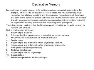
Systems mediating declaractive memory functions Evidence for the importance of hippocampal and parahippocampal systems in declarative memory in human: “H.M. appears to have a complete loss of memory for events [anterograde amnesia] subsequent to bilateral medial temporal lobe resection 19 months before, together with a partial retrograde amnesia for the 3 months leading up to his operation, but early memories are seemingly normal and there is no impairment of personality or general intelligence” in Scoville and Milner J. Neurol. Neurosurg. Psychiatry 1957, 20: 11-21. “Every day is alone, whatever enjoyment I’ve had and whatever sorrow I’ve had” (H.M.)
MRI images of patient H.M.: Bilateral anterior lobectomy.
The delayed non-match to sample task (Mishkin and Appenzeller 1987) This task served to investigate which structures of the medial temporal lobe are critical for declarative memory (see Larry Squire Box 19.3 “Neuroscience” Bear, Connors and Paradiso 1996).
Monkeys with lesions of: H+: hippocampal formation, parahippocampal cortex. H++: idem and and perirhinal cortex. Zola-Morgan et al 1993
Performance of the delayed non-matching to sample task for normal (N), and lesioned monkeys (H+ and H++).
relevance septal area Hippocampal memory systems Association areas: frontal, temporal, parietal, occipital Hippocampal formation sensory map Postrhinal & perirhinal cortices Subiculum sensory map & novelty detection CA1 Medial & lateral entorhinal cortices DG/CA3 Lopes da Silva et al 2000
Distinct memory systems mediating declarative, emotional and proceduralmemory functions • Main anatomic memory systems: • Declarative memory; • Emotional memory; fear conditioning; • Motor conditioning; • Skills and Priming.
MTL: (para)hippocampal cortex Taxonomy of memory Declarative (explicit) Procedural (implicit) Episodic (working) Semantic (reference) Skills Priming Conditioning Non-associative Emotional responses Motor responses MTL: (para)hippocampal cortex Reflex paths Amygdala Striatum Cortex Amygdala Cerebellum Diencephalon (Adapted from L.R. Squire: “Memory and the Brain”, 1987)
Sagittal view of the brain showing the location of the amygdaloid complex of nuclei in the temporal lobe. Coronal section through the forebrain at the leveel of the amygdala. (Purves et al “Neuroscience”, 2nd edition, 2001, Box B, p.634)
Neural circuits involved in conditioned emotional responses, specifically fear conditioning. (LeDoux, J.E. Emotion: clues from the brain. Annu. Rev. Psychol. 1995, 46: 209-235).
Simple fear conditioned task, aversive classical conditioning: Temporal pairing of a neutral stimulus (CS=sound) with an aversive event (US) (foot shock). Conditioned response: freezing with autonomic and hormonal components. LeDoux, Annu. Rev. Psychol. 1995, 46: 209-235). .
Pathways that mediate the association of auditory and aversive stimuli. (Purves et al “Neuroscience” 2nd edition, 2001, p. 637)
Model of associative learning in the amygdala relevant to emotional conditioning (Rolls, 1999).
Conditioned fear-induced changes in the receptive field (RF) properties of single neurons in the amygdala. Neuron’s best response to a tone frequency: BF. BF determined before conditioning = Pre; After conditioning = Post. After conditioning the BF of the neuron shifted to the frequency of the CS tone.
Distinct memory systems mediating declarative, emotional and proceduralmemory functions • Main anatomic memory systems: • Declarative memory; • Emotional memory; fear conditioning; • Motor conditioning; • Skills and Priming.
MTL: (para)hippocampal cortex Taxonomy of Memory Declarative (explicit) Procedural (implicit) Episodic (working) Semantic (reference) Skills Priming Conditioning Non-associative Emotional responses Motor responses MTL: (para)hippocampal cortex Reflex paths Cerebellum Cerebellum Striatum Cortex Amygdala & Diencephalon (Adapted from L.R. Squire: “Memory and the Brain”, 1987)
Classical conditioning as it develops in the course of training: The response is the eyelid closure shown as an upward deflection of the trace. US: air puff to the cornea. CS: neutral tone.
The Cerebellum plays an essential role in classical conditioning of motor resposnses such as in the eye blink reflex, elicited by a corneal air puff.
Scheme of the eye blink conditioning circuit. US (air puff) path: N V (trigeminus) cranial nerve, inferior olive (IO) climbing fibers (CF), Purkinje cells. CS (tone) path: Cochlear nerve, pontine nuclei (PN), mossy fibers (MF), granule cells (GR), parallel fibers (PF). Efferent path: Purkinje cells, Interpositus nucleus (Int), red nucleus (RN), motor nuclei (VI and VII cranial nerves).
Responses of cerebellar interpositus neurons in eye blink conditioning: before CS-US pairing and at days 1 and 2 after pairing. Total trace duration: 750 ms; CS – US onset interval: 250 ms. (From McCormick and Thompson , Science 1984)
Memory systems Are the hippocampal memory systems also engaged in this form of classical conditioning (eye blink reflex)? Patients with temporal lobe anterograde amnesia are able to learn the acquisition of the eye blink conditioned response, but cannot recall the learning experience. However this ability depends on the complexity of the conditioned task. If the task is to blink to tone if, and only if, preceded by light, they show also impairment.
Distinct memory systems mediating declarative, emotional and proceduralmemory functions • Main anatomic memory systems: • Declarative memory; • Emotional memory; fear conditioning; • Motor conditioning; • Skills and Priming.
Regions exhibiting skill learning and priming -related changes in fMRI activity: Increases: R Cerebellum (-21); L Inferior Frontal (+33); L Inferior Temporal (-12); Anterior Cingulate; Tail of Caudate (+15); Decreases: L Cerebellum; (-21) L Hippocampus (-18). Poldrack and Gabrieli, Brain 2001, 124:67-82
Memory systems All regions exhibiting significant learning-related changes also exhibited increased repetition priming effects, suggesting common neural substrates for priming and skill learning. The results of the study of Poldrack and Gabrieli indicate the importance of striato-frontal neural networks for the memory for skills.
Distinct memory systems mediating declarative, emotional and proceduralmemory functions • Main anatomic memory systems: • Declarative memory; • Emotional memory; fear conditioning; • Motor conditioning; • Skills and Priming.
Memory systems One region that did not emerge specifically in the previous analysis is the prefrontal cortex. Is this region also relevant as a part of memory systems? Which?
The prefrontal cortex has been suggested to be necessary for strategic memory processes and for retrieval. These strategic processes may facilitate episodic learning. This study examined strategic memory with PET using a verbaal encoding paradigm that manipulated semantic organization in differenty conditions.
Three tasks: • Spontaneous – words were related in four semantic categories but subjects were not aware of this beforehand; • Directed: in this condition the subjects were instructed that there were 4 semantic categories; • Unrelated: words did not share any semantic relationship. Regions of activation represent voxels where rCBF increased in a graded fashion: directed> spontaneous>unrelated: L lateral prefrontal cortex; L Inferior frontal gyrus.
Memory systems Understanding the neurobiology of memory processes involves not only the identification of the memory systems that are anatomically involved in the storage of information but also the equally important question of how information is stored. Hebb pointed out already in 1949 that memories can result from subtle changes at the level of synapses. This basic assumption has led to search for the physical basis of memories in synaptic plasticity.
“The assumption, in brief, is that a growth process accompanying synaptic activity makes the synapse more readily traversed.” According to this hypothesis, “an intimate relationship is postulated between reverberatory action and structural changes at the synapse, implying a dual trace mechanism”.
Hebb hypothesis led to the experimental strategy that revealed the existence of long-term potentiation by Bliss and Lomo in 1966 – 1968.
Typical set-up to demonstrate the existence of LTP in the synapses that the Schaffer collaterals of CA3 neurons make with the dendrites of CA1 neurons of the Hippocampus. (Malinow et al 1989)
Varieties of synaptic plasticity: Neurons receive either strong (S) or weak (w) stimuli. Postsynaptic potentials before inducing stimulation are given as solid lines; after stimulation as dotted lines.
Long-term synaptic depression (LTD) caused by low-frequency stimulation (1Hz). (Mulkey and Malenka 1992)
Main events leading to LTP or to LTD. If both LTP and LTD are triggered by [Ca2+]i, how are the induction processes different? The hypothesis is that high [Ca2+]i (>5mM) activates a protein kinase leading to LTP, while if [Ca2+]i does not increase more than 5mM a phophatase is activated causing LTD. (Zigmond et al “Fundamental Neuroscience” 1999, p. 1444)
According to Shi et al LTP and LTD can arise from the rapid redistribution (insertion in LTP or retrieval in LTD) of Glutamate AMPA receptors after synaptic NMDA receptor activation depending on the stimulus properties. (Science 1999, 284: 1811-16)