Download

1 / 15
150 likes | 163 Views
Bacterial Structure (Lab 3 ). Rana Alqusumi. Cell wall. Cell membrane. Cytoplasm. Nuclear material. Essential Bacterial Structures. Capsule. Flagella. Pili. Fimbriae. Spore. Particular Bacterial Structures. Bacterial Structure (Capsule). Glycocalyx :

E N D
Bacterial Structure (Lab 3) Rana Alqusumi
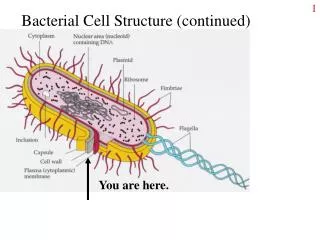
Cell wall. • Cell membrane. • Cytoplasm. • Nuclear material. EssentialBacterial Structures
Capsule. • Flagella. • Pili. • Fimbriae. • Spore. ParticularBacterial Structures
Glycocalyx : some extracellular material secreted by many bacterial cells in the form of: • capsule : attached tightly to the bacterium and has definite boundaries. • slime layer : loosely associated with the bacterium and can be easily washed of. Cell Envelope
A capsule is a gelatinous outer layer that is secreted by the cell and that surrounds and adheres to the cell wall. It is not common to all organisms. • Cells that have a heavy capsule are generally virulent and capable of producing disease, since the structure protects bacteria against the normal phagocytic activities of host cells. Capsule
The capsule stain uses two reagents: • Primary Stain: Crystal Violet (1% aqueous) A violet stain is applied to a non–heat-fixed smear. At this point, the cell and the capsular material will take on the dark color. Capsule Staining
Decolorizing Agent: • Copper Sulfate (20%) Because the capsule is nonionic, unlike the bacterial cell, the primary stain adheres to the capsule but does not bind to it. • In the capsule staining method, copper sulfate is used as a decolorizing agent rather than water. • The copper sulfate washes the purple primary stain out of the capsular material without removing the stain bound to the cell wall. At the same time, the decolorized capsule absorbs the copper sulfate, and the capsule will now appear blue in contrast to the deep purple color of the cell. Capsule Staining
Materials: • Culture: 48-hour-old cultures of bacteria. • Reagents: 1% crystal violet and 20% copper sulfate. • Equipment: • Bunsen burner, • Inoculating loop or needle, • Glass slides. • Microscope. Capsule Stain(Anthony Method)
Procedure: • Steps 1–5 are pictured in Figure 13.5. • Obtain one clean glass slide. Place several drops of crystal violet stain on the slide. • Using aseptic technique, add three loopfuls of a culture to the stain and gently mix with the inoculating loop. • With a clean glass slide, spread the mixture over the entire surface of the slide to create a very thin smear. Let stand for 5 to 7 minutes. Allow smears to air-dry. Note: Do not heat fix. • Wash smears with 20% copper sulfate solution. • Gently blot dry and examine under oil immersion. Capsule Stain(Anthony Method)
James G. Cappuccino, Natalie Sherman. 2014. Microbiology a laboratory manual. 10th ed. • https://fac.ksu.edu.sa/sites/default/files/362mic-lab3.pdf Reference