Download
1 / 1
30 likes | 222 Views
Fibroblast Derived Cytokines Improve Skeletal Muscle Differentiation in a Strain Dependent Manner Michael Hicks 2 , Thanh Cao 1 , David Campbell 1 , and Paul Standley 1 1 University of Arizona-Phoenix, AZ; 2 Arizona State University-Tempe, AZ. Co-culture System.

E N D
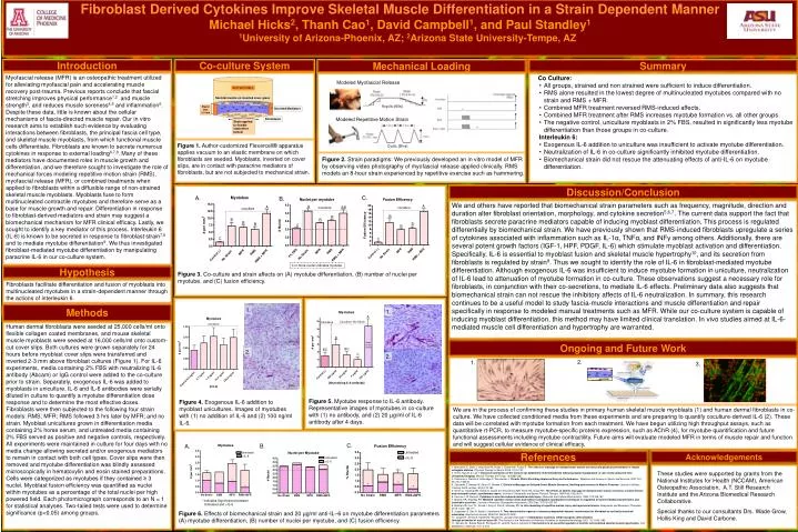
Fibroblast Derived Cytokines Improve Skeletal Muscle Differentiation in a Strain Dependent Manner Michael Hicks2, Thanh Cao1, David Campbell1, and Paul Standley1 1University of Arizona-Phoenix, AZ; 2Arizona State University-Tempe, AZ Co-culture System Modeled Repetitive Motion Strain Cyclic (8hrs) Modeled Myofascial Release Acyclic (60s) Hypothesis Fibroblasts facilitate differentiation and fusion of myoblasts into multinucleated myotubes in a strain-dependent manner through the actions of interleukin 6. 1. 2. 1. Interleukin-6 Summary Introduction Mechanical Loading Myofascial release (MFR) is an osteopathic treatment utilized for alleviating myofascial pain and accelerating muscle recovery post-trauma. Previous reports conclude that fascial stretching improves physical performance1,2 and muscle strength3, and reduces muscle soreness4,5 and inflammation5. Despite these data, little is known about the cellular mechanisms of fascia-directed muscle repair. Our in vitro research aims to establish such evidence by evaluating interactions between fibroblasts, the principal fascia cell type, and skeletal muscle myoblasts, from which functional muscle cells differentiate. Fibroblasts are known to secrete numerous cytokines in response to external loading6,7,8. Many of these mediators have documented roles in muscle growth and differentiation, and we therefore sought to investigate the role of mechanical forces modeling repetitive motion strain (RMS), myofascial release (MFR), or combined treatments when applied to fibroblasts within a diffusible range of non-strained skeletal muscle myoblasts. Myoblasts fuse to form multinucleated contractile myotubes and therefore serve as a base for muscle growth and repair. Differentiation in response to fibroblast-derived mediators and strain may suggest a biomechanical mechanism for MFR clinical efficacy. Lastly, we sought to identify a key mediator of this process. Interleukin 6 (IL-6) is known to be secreted in response to fibroblast strain7,8 and to mediate myotube differentiation9. We thus investigated fibroblast-mediated myotube differentiation by manipulating paracrine IL-6 in our co-culture system. • Co Culture: • All groups, strained and non strained were sufficient to induce differentiation. • RMS alone resulted in the lowest degree of multinucleated myotubes compared with no strain and RMS + MFR. • Combined MFR treatment reversed RMS-induced affects. • Combined MFR treatment after RMS increases myotube formation vs. all other groups. • The negative control, uniculture myoblasts in 2% FBS, resulted in significantly less myotube differentiation than those groups in co-culture. • Interleukin 6: • Exogenous IL-6 addition to uniculture was insufficient to activate myotube differentiation. • Neutralization of IL-6 in co-culture significantly inhibited myotube differentiation. • Biomechanical strain did not rescue the attenuating effects of anti-IL-6 on myotube differentiation. BIOFLEX WELL Skeletal muscle on inverted cover glass Plastic Rim 2.5 mm Secreted Mediators Fibroblasts Strain applied to flexible elastomere bottom Figure 1. Author-customized Flexercell® apparatus applies vacuum to an elastic membrane on which fibroblasts are seeded. Myoblasts, inverted on cover slips, are in contact with paracrine mediators of fibroblasts, but are not subjected to mechanical strain. Figure 2. Strain paradigms: We previously developed an in vitro model of MFR by observing video photography of myofascial release applied clinically. RMS models an 8-hour strain experienced by repetitive exercise such as hammering. Discussion/Conclusion C. A. B. We and others have reported that biomechanical strain parameters such as frequency, magnitude, direction and duration alter fibroblast orientation, morphology, and cytokine secretion5,6,7. The current data support the fact that fibroblasts secrete paracrine mediators capable of inducing myoblast differentiation. This process is regulated differentially by biomechanical strain. We have previously shown that RMS-induced fibroblasts upregulate a series of cytokines associated with inflammation such as IL-1α, TNFα, and INFγ among others. Additionally, there are several potent growth factors (IGF-1, HPF, PDGF, IL-6) which stimulate myoblast activation and differentiation. Specifically, IL-6 is essential to myoblast fusion and skeletal muscle hypertrophy10, and its secretion from fibroblasts is regulated by strain8. Thus we sought to identify the role of IL-6 in fibroblast-mediated myotube differentiation. Although exogenous IL-6 was insufficient to induce myotube formation in uniculture, neutralization of IL-6 lead to attenuation of myotube formation in co-culture. These observations suggest a necessary role for fibroblasts, in conjunction with their co-secretions, to mediate IL-6 effects. Preliminary data also suggests that biomechanical strain can not rescue the inhibitory affects of IL-6 neutralization. In summary, this research continues to be a useful model to study fascia-muscle interactions and muscle differentiation and repair specifically in response to modeled manual treatments such as MFR. While our co-culture system is capable of inducing myoblast differentiation, this method may have limited clinical translation. In vivo studies aimed at IL-6-mediated muscle cell differentiation and hypertrophy are warranted. Figure 3. Co-culture and strain affects on (A) myotube differentiation, (B) number of nuclei per myotube, and (C) fusion efficiency. Methods 1. Human dermal fibroblasts were seeded at 25,000 cells/ml onto flexible collagen coated membranes, and mouse skeletal muscle myoblasts were seeded at 16,000 cells/ml onto custom-cut cover slips. Both cultures were grown separately for 24 hours before myoblast cover slips were transferred and inverted 2-3 mm above fibroblast cultures (Figure 1). For IL-6 experiments, media containing 2% FBS with neutralizing IL-6 antibody (Abcam) or IgG control were added to the co-culture prior to strain. Separately, exogenous IL-6 was added to myoblasts in uniculture. IL-6 and IL-6 antibodies were serially diluted in culture to quantify a myotube differentiation dose response and to determine the most effective doses. Fibroblasts were then subjected to the following four strain models: RMS; MFR; RMS followed 3 hrs later by MFR; and no strain. Myoblast unicultures grown in differentiation media containing 2% horse serum, and untreated media containing 2% FBS served as positive and negative controls, respectively. All experiments were maintained in culture for four days with no media change allowing secreted and/or exogenous mediators to remain in contact with both cell types. Cover slips were then removed and myotube differentiation was blindly assessed microscopically in hematoxylin and eosin stained preparations. Cells were categorized as myotubes if they contained ≥ 3 nuclei. Myoblast fusion efficiency was quantified as nuclei within myotubes as a percentage of the total nuclei per high powered field. Each photomicrograph corresponds to an N = 1 for statistical analyses. Two-tailed tests were used to determine significance (p<0.05) among groups. Ongoing and Future Work 2. 2. 3. Figure 5. Myotube response to IL-6 antibody. Representative images of myotubes in co-culture with (1) no antibody, and (2) 20 µg/ml of IL-6 antibody after 4 days. Figure 4. Exogenous IL-6additionto myoblast unicultures. Images of myotubes with (1) no addition of IL-6 and (2) 100 ng/ml IL-6. We are in the process of confirming these studies in primary human skeletal muscle myoblasts (1) and human dermal fibroblasts in co-culture. We have collected conditioned media from these experiments and are preparing to quantify coculture-derived IL-6 (2). These data will be correlated with myotube formation from each treatment. We have begun utilizing high throughput assays, such as quantitative rt-PCR, to measure myotube-specific proteins expression, such as AChR (4), for myotube quantification and future functional assessments including myotube contractility. Future aims will evaluate modeled MFR in terms of muscle repair and function and will suggest cellular evidence of clinical efficacy. C. B. A. Acknowledgements References 1. Mancinelli D, Davis L, Aboulhosn M, Brady J, Eisenhofer, Foutty S. The effects of massage on delayed onset muscle soreness and physical performance in female collegiate athletes. Physical Therapy in Sports. 2006; 7(1)5-13C. 2. Gill N, Teyhen D, Lee I. Improved contraction of the transversus abdominis (TrA) immediately following spinal manipulation: A case study using real-time ultrasound imaging.Manual Therapy, 2007 Aug; 12(3)280-285 3. Kokkonen J, Nelson A, Elderedge C, Winchester J. Chronic Static Stretching Improves Exercise Performance.Medicine and Science in Sports and Exercise. 2007 Oct; 39 (10): 1825-31 4. Zainuddin Z, Newton M, Sacco P, Nosaka K. Effects of Massage on Delayed-Onset Muscle Soreness, Swelling and recovery of Muscle Function.Journal of Athletic Training. 2005 Jul-Sep; 40(3):174-180. 5. Smith LL, Keating MN, Holber D, Spratt DJ, McCammon MR, Smith SS, Istreal RG. The effects of athletic massage on delayed onset muscle soreness, creatine kinase, and neutrophil count: a preliminary report.Journal of Orthopedic and Sports Physical Therapy. 1994 Feb; 19(2):93-9 6. Cannon J, St Pierre B. Cytokines in exertion-induced skeletal muscle injury. Molecular and Cellular Biochemistry. 1998; 179:159-167. 7. Meltzer KR, Standley PR. Modeled repetitive motion strain and indirect osteopathic manipulative techniques in regulation of human fibroblast proliferation and interleukin secretion. Journal of American Osteopathic Association. 2007 Dec; 107(12):527-36 8. Melzter KR, Cao TV, Schad J, King H, Stoll S, Standley PR. In vitro modeling of repetitive motion injury and myofascial release.Bodyworks and Movement Therapies. 2010; 14(2): 162-171. 9. Jungbauer S, Gao H, Spatz J, Kemkemer R. Two characteristic regimes in frequency-dependent dynamic reorientation for fibroblast on cyclically stretched substrates. Biophysical Journal, 2008 Oct; (95)3470-3478 10. Langen R, Schols A, Kelders M, Wouters E,Janssen-Heininger Y. Inflammatory cytokines inhibit myogenic differentiation through activation of nuclear factor-kB. The Journal of the Federation of American Societies for experimental biology.2001; 15: 1169-1180 11. Serrano AL, Baeza-Raja B, Perdiguero E, Jardi M, Munoz-Canoves P. Interluekin-6 is an essential regulator of satellite cell-mediated skeletal muscle hypertrophy.Cell Metabolism. 2008 Jan; 7(1): 33-44 These studies were supported by grants from the National Institutes for Health (NCCAM), American Osteopathic Association, A.T. Still Research Institute and the Arizona Biomedical Research Collaborative. Special thanks to our consultants Drs. Wade Grow, Hollis King and David Carbone. Figure 6. Effects of biomechanical strain and 20 µg/ml anti-IL–6 on myotube differentiation parameters (A) myotube differentiation, (B) number of nuclei per myotube, and (C) fusion efficiency. .