Download
1 / 1
10 likes | 131 Views
RECENT OPTICAL AND SEM CHARACTERIZATION OF GENESIS SOLAR WIND CONCENTRATOR DIAMOND-ON-SILICON COLLECTOR.

E N D
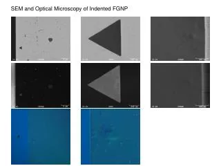
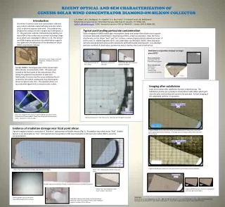
RECENT OPTICAL AND SEM CHARACTERIZATION OF GENESIS SOLAR WIND CONCENTRATOR DIAMOND-ON-SILICON COLLECTOR J. H. Allton1 , M. C. Rodriguez2, P. J. Burkett3, D. K. Ross3 and C. P. Gonzalez3 and K. M. McNamara1, 1NASA/Johnson Space Center, 2101 NASA Parkway, Mail Code KT, Houston, TX 77058, USA, Judith.h.allton@nasa.gov, 2 ESGC Geocontrol Systems- ESCG at NASA/JSC, 3 Jacobs- ESCG at NASA /JSC. Introduction One of the 4 Genesis solar wind concentrator collectors was a silicon substrate coated with diamond-like carbon (DLC) in which to capture solar wind. This material was designed for analysis of solar nitrogen and noble gases [1, 2]. This particular collector fractured during landing, but about 80% of the surface was recovered, including a large piece which was subdivided in 2012 [3, 4, 5]. The optical and SEM imaging and analysis described below supports the subdivision and allocation of the diamond-on-silicon (DOS) concentrator collector. Typical post-landing particulate contamination Optical imaging of 60000 reveals glass microsphere shards and carbon fibers (both return capsule structural materials), dried droplets, miscellaneous fibers, smears and particles. Near the frame attachments, on the silicon “heel” and “toe” surfaces, smears of gold-colored material are noted. A brief survey of related fragment, 60737, was undertaken via SEM (JEOL 7600F). Most abundant particles were gold (with nickel and/or aluminum) and GaZnAl-oxide bearing paint. Less abundant particles consisted of silicate glass, germanium metal, stainless steel, and terrestrial soil. SEM debris composition analyses on target piece 60737 Main contaminants: gold metal ± Ni ± Al GaZnAlO bearing paint Minor contaminants silicate glass Ge – metal stainless steel terrestrial soil particles Diamond-on-silicon target recovered fragments. Quadrant is 3 mm on edge. Crash-derived smear on DLC surface. DLC layers at corner of clip mark. Hole in DLC film with “radiation texture” on silicon. Sample 60000 is the largest piece of the concentrator DOS collector, as recovered in Utah. This piece was located at the focal point of the concentrator; thus, having the greatest concentration of solar ions. Additionally, this piece had two areas inadvertently not covered by the carbon coating and, thus exposing the silicon to capture solar ions. The exposed silicon, set up a potential opportunity to measure solar carbon. Imaging after subdivision Solar wind surface after subdivision by laser scribe/cleaving. The subdivision process [4, 5] produces silicon/silicon oxide debris which gets onto the solar wind surface and needs to be removed. Further imaging of the subdivided surfaces is in progress. False color image of DOS collector fragment 60000 mounted in frame and location of frame shadow. Green areas show solar-wind-exposed silicon. Quadrant is 3 mm on edge. Debris and smears on “toe” silicon area. Gold may be from gold cross frame. Evidence of irradiation damage near focal point silicon Optical imaging revealed a correlation of “blueness” and presence of bubble texture (Fig. 5). No gradient was noted across “heel”. Bubble texture is not observable on “toe”. Estimated proton flux gradient is 20X near focal point vs 6X near outer radius (Wiens, personal communication). Fragment 60000 color-enhanced to show increased blueness of silicon areas near focal point, presumably due to irradiation. The bright areas are silicon, called “heel” and “toe” of sock shape. Fragment 60000 post subdivision contrast stretched to show debris from subdivision. Silicon “heel” showing frame shadow and gold-colored smears. Silicon “toe” showing gold-colored smear and debris. Bubble texture on blue areas of silicon Bubble features are micrometer diameter. Silicon “toe” area exposed to solar wind is shown is dark blue. Fragment 60000 just prior to last cut, removal of some laser-generated debris. References: [1] Nordholt J. E. et al (2003) Space Sci. Rev., 105, 561-599. [2] Jurewicz A. J. G. et al. (2003) Spa. Sci. Rev., 105, 535-560 (2002), [3] Rodriguez et al (2009) LPS XL, Abst. #1337, [4] Burkett, P. J. et al. (2013), LPS XLIV [5] Lauer H. V. et al (2013) LPS XLIV. Texture gradient across solar wind exposure boundary. FOV is 220 µm. Anomalous feature under clamp area during carbon deposition. Glass microsphere shard on left. “Radiation texture” on silicon. FOV is 220 µm.