Download
1 / 12
120 likes | 292 Views
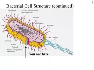
Phage head. Tail sheath. Tail fiber. DNA. 100 nm. Bacterial cell. EXPERIMENT. Empty protein shell. Radioactivity (phage protein) in liquid. Radioactive protein. Phage. Bacterial cell. DNA. Batch 1: radioactive sulfur ( 35 S). Phage DNA. Centrifuge.

E N D
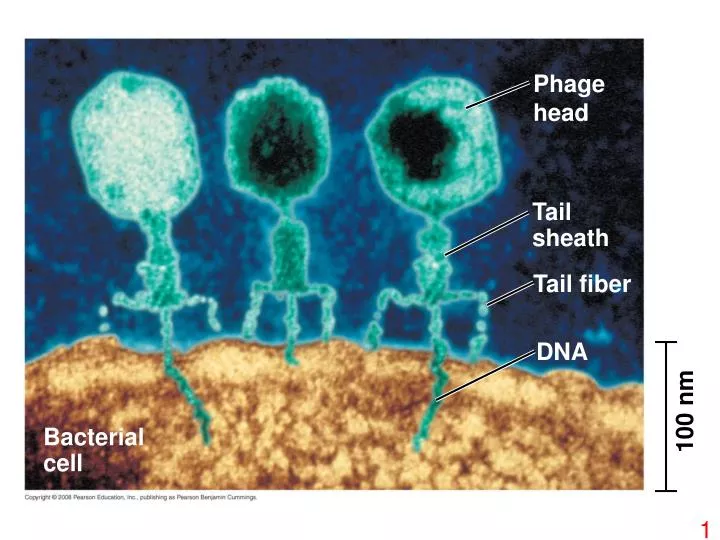
Phage head Tail sheath Tail fiber DNA 100 nm Bacterial cell
EXPERIMENT Empty protein shell Radioactivity (phage protein) in liquid Radioactive protein Phage Bacterial cell DNA Batch 1: radioactive sulfur (35S) Phage DNA Centrifuge Pellet (bacterial cells and contents) Radioactive DNA Batch 2: radioactive phosphorus (32P) Centrifuge Radioactivity (phage DNA) in pellet Pellet
Nitrogenous bases Sugar–phosphate backbone 5 end Thymine (T) Adenine (A) Cytosine (C) DNA nucleotide Phosphate Sugar (deoxyribose) 3 end Guanine (G)
(b) Franklin’s X-ray diffraction photograph of DNA (a) Rosalind Franklin
5 end Hydrogen bond 3 end 1 nm 3.4 nm 3 end 0.34 nm 5 end (c) Space-filling model (a) Key features of DNA structure (b) Partial chemical structure
Purine + purine: too wide Pyrimidine + pyrimidine: too narrow Purine + pyrimidine: width consistent with X-ray data
Adenine (A) Thymine (T) Cytosine (C) Guanine (G)
A T A T A T A T C G C G C G C G A T A T A A T T T A T A T T A A C C G C G C G G (c) “Daughter” DNA molecules, each consisting of one parental strand and one new strand (b) Separation of strands (a) Parent molecule
Origin of replication Parental (template) strand Daughter (new) strand Replication fork Double- stranded DNA molecule Replication bubble 0.5 µm Two daughter DNA molecules (a) Origins of replication in E. coli Origin of replication Double-stranded DNA molecule Parental (template) strand Daughter (new) strand 0.25 µm Replication fork Bubble Two daughter DNA molecules (b) Origins of replication in eukaryotes
New strand 5 end Template strand 3 end 5 end 3 end Sugar T A A T Base Phosphate C G C G G C G C DNA polymerase 3 end A A T T 3 end Pyrophosphate C C Nucleoside triphosphate 5 end 5 end
Nucleosome (10 nm in diameter) DNA double helix (2 nm in diameter) H1 Histone tail Histones DNA, the double helix Histones Nucleosomes, or “beads on a string” (10-nm fiber)
Chromatid (700 nm) 30-nm fiber Loops Scaffold 300-nm fiber Replicated chromosome (1,400 nm) 30-nm fiber Looped domains (300-nm fiber) Metaphase chromosome