Download
1 / 63
920 likes | 3.01k Views
Heart Disease Braunwald. CV R4 李威廷醫師 Supervisor: 劉秉彥醫師. Pathophysiology of Heart Failure. Myocardial failure: myocardial infarction, acute myocarditis Heart failure: myocardial failure, acute AR, constrictive pericarditis Circulatory failure:

E N D
Heart Disease Braunwald CV R4 李威廷醫師 Supervisor: 劉秉彥醫師 Pathophysiology of Heart Failure
Myocardial failure: myocardial infarction, acute myocarditis Heart failure: myocardial failure, acute AR, constrictive pericarditis Circulatory failure: heart failure, hypovolemic shock, septic shock
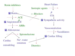
Adaptive mechanisms • Frank-starling mechanism: preload (short-term) • Neuroendocrine system: norepinephrine (short-term) • Myocardial remodeling: with or without chamber dilatation (chronic or long-term)
Vascular redistribution • Increase vasoconstrictor activity sympathetic nervous system renin-angiotensin system endothelin system keep adequate oxygen to vital organs (brain, heart) • Endothelial dysfunction ischemic- and exercise- induced vasodilation attenuated L-arginine, NO
Peripheral hypoperfusion • Non-vital organs: Anaerobic metabolism, lactic acidosis • Skin: cold, dry turger • Muscle: weakness • Kidney: sodium and nitrogen retention • Gut: decreased GI motility, GI upset • Liver: hepatic dysfunction
Chronic myocardial remodeling (1) • Pressure overload: increase ventricular wall thickness (concentric hypertrophy) (isometric) • Volume overload: mild increase ventricular wall thickness (eccentric hypertrophy)(counterbalance the increased radius) (isotonic)
Molecular mechanism of myocardial remodeling and failure(Myocyte Loss) • Necrosis: deprived of oxygen or energy loss of membrane integrity influx of ECF cellular swelling release of proteolytic enzymes cellular disruption (subendocardial area) (acute myocarditis, DCM, myocardial infarction) • Apoptosis: specific genetic program molecular cascade of degradation of nuclear DNA (angiotensin II, NO, inflammatory cytokines, mechanical strain)
Molecular mechanism of myocardial remodeling and failure(Excitation-Contraction coupling alternation) • Ca: contraction and relaxation; • Force-frequency relationship: contractile force decrease, rates of stimulation increase • Sarcoplasmic reticulum Ca-ATPase & phospholamban: Ca reuptake system (SERCA2) • Na-Ca exchanger: removal Ca in diastole • Ca free channel, voltage-dependent Ca channel: • Calsequestrin: major Ca binding protein in SR
Molecular mechanism of myocardial remodeling and failure(Contractile apparatus alternation) • Reduction of myosin ATPase activity: qualitative, quantitative • Myosin isoform changes: fetal and neonatal form • Altered regulatory proteins: troponin-T1 T2
Molecular mechanism of myocardial remodeling and failure(Matrix alternation) • Regulation of interstitial collagen: • Collagen strut depletion: • Interstitial matrix accumulation:
Pathophysiology of diastolic heart failure • Altered ventricular relaxation relaxation: inactivation of contraction isovolemic relaxation and early ventricular filling SERCA2 and calcium pump ischemia, elevated afterload • Altered ventricular filling wall stiffness diastolic asynergy: regional abnormal relaxation diastolic asynchony: no relaxation ischemia, pericardial disease
Neurohormonal, autocrine, & paracrine adjustments (1) • Response to inadequate arterial volumesystolic HF • Adrenergic • Renin-angiotensin-aldosterone • Vasopressin and endothelin • Atrial natriuretic peptide (ANP)
Neuroendocrine, autocrine, & paracrine adjustments (2) • Autonomic nervous system Increased sympathetic activity: bloodnorepinephrine, abnormalbaroreflex (?) Decreased parasympathetic activity: Cardiac norepinephrine depletion: not clear Beta1-adrenergic receptor density decrease, G protein: local NE concentration, beta1-antagonist Gs (stimulation), Gi (inhibition): adenynyl cyclase
Neuroendocrine, autocrine, & paracrine adjustments (3) • Renin-angiotensin system (RAS) Juxtaglomerular beta1-receptor: renin Renal vascular baroreceptor: renin Adrenergic nervous system: NE • Tissue RAS: 90—99% Early activation than blood RAS • Angiotensin receptor: AT1, AT2
Neuroendocrine, autocrine, & paracrine adjustments (4) • Arginine vasopressin (AVP): V1 receptor, cathecholamine • Natriuretic peptides: Atrial-NP, Brain-NP, C-NP • Endothelin: pulmonary vasoconstrictor, Killip stage • Inflammatory cytokine: TNF-a, IL-1b; Ca & myocardium • Nitric oxide: inhibit inflammatory cytokines, apoptosis • Oxidative stress: (animal study)
Clinical Aspects of heart failure: high-output heart failure; pulmonary edema CV R4 李威廷醫師 Supervisor: 劉秉彥醫師 Nov 6th, 2003
Backward heart failure hypothesis • Right ventricular failure as a sequence of left ventricular failure • Ventricle failed to discharge its contents • Increase LVEDP • Blood accumulation and pressure arises in atrium and venous system • The atrium contracts more vigorously • Venous and capillary pressure arise • Fluid transudation from capillary bed to interstitial space
Forward heart failure hypothesis • Reduced cardiac output, then diminished perfusion of vital organs • Sodium and water retention • Increased extracellular fluid • Congestion of organs and tissues • Massive myocardial infarction both forward and backward heart failure hypothesis
Low output versus high output heart failure • Low output heart failure congenital, valvular, rheumatic, hypertensive, coronary, and cardiomyopathic heart failure • High output heart failure thyrotoxicosis, arteriovenous fistula, anemia, beriberi disease, Paget’s disease of bone
Causes of heart failure • Underlying disease: structural, vessel, or valvular • Fundamental causes: increased hemodynamic burden or reduction in oxygen delivery • Precipitating causes: specific causes or incidents (avoidance of a precipitating cause can prevent heart failure)
Precipitating causes of heart failure (1) • Inapposite reduction of therapy: Na, water, diuretics • Arrhythmia: tachycardia, bradycardia, abnormal conduction • Myocardial ischemia or infarction • Systemic infection: esp. pulmonary • Pulmonary embolism • Physical, emotional, and environmental stress • Cardiac infection and inflammation: myocarditis • Development of an unrelated illness: ARF
Precipitating causes of heart failure (2) • Cardiac depressant of salt-retaining drug: beta- blocker, isoptin, diltiazm, doxorubicin, cyclophosphamide • Cardiac toxins: alcohol, cocaine • High-output status: valvular heart disease, anemia, pregnancy • A second form of heart failure: HCVD + AMI
Symptoms of heart failure • Respiratory distress • Reduced exercise capacity • Physical findings • Laboratory findings • CXR
Respiratory distress • Exertional dyspnea: the degreeof activity necessary tp induce the symptom • Orthopnea: dyspnea at recumbent position, relieving by elevation of the head with pillows (not-specific) • Paroxysmal nocturnal dyspnea: interstitial pulmonary edema bronchospasm wheezing (cardiac asthma) • Dyspnea at rest • Acute pulmonary edema
Pulmonary & Cardiac Dyspnea • Cough v.s. sitting up relief • Smoking history • Bronchodilator agent v.s. diuretics
Reduced exercise capacity (1) • Mechanism Pulmonary vascular congestion Inadequate blood flow to exercise muscle Deconditioning of skeletal and respiratory muscles Attenuated peripheral blood vessel resistance Abnormal skeletal metabolism Patient anxiety
Reduced exercise capacity (2) • Exercise testing Maximal exercise capacity: treadmill test (total oxygen uptakes) reflecting central hemodynamic response adequacy Submaximal exercise capacity: 6-minute walk test reflecting regulation of blood flow to the skeletal muscle • Other organs symptoms Brain (confusion, insomnia), urinary tract (nocturia)
Reduced exercise capacity (3) • Functional classification: (New York Heart Association) I: no limitation II: slight limitation III: marked limitation IV: inability for any work without discomfort • Quality of life: reduce symptoms, prolong survival, and improve quality of life
Physical findings • General appearance: orthopnea, malnutrition, cyanosis • Increased adrenergic activity: pallor, cold, diaphoresis • Pulmonary rales • Systemic venous hypertension: JVE, Kussmaul’s sign • Hepatojugular reflux: right side heart • Congestive hepatomegaly • Edema: extracelluar fluid gain >4L • Pleural effusion: R’t ± L’t • Ascites: long-term systemic venous hypertension