Download
1 / 6
230 likes | 1.84k Views
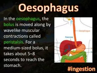
Oesophagus. Normal . What radiologic studies can be used to image the esophagus ? Investigations: Barium Swallow. CT and MRI Endoscopic ultrasound Questions: 1. What is this investigation ? 2. Describe the radiological findings. 3. What is your diagnosis ?. Oesophageal Varices.

E N D
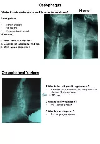
Oesophagus Normal What radiologic studies can be used to image the esophagus ? Investigations: • Barium Swallow. • CT and MRI • Endoscopic ultrasound Questions: 1. What is this investigation ? 2. Describe the radiological findings. 3. What is your diagnosis ? OesophagealVarices • 1. What is the radiographic appearance ? • There are multiple submucosal filling defects in a barium filled esophagus • in AP view. • 2. What is this investigation ? • Ans: Barium Swallow • 3. What is your diagnosis ? • Ans: esophageal varices.
1. What is the investigation ? • Barium Swallow . • 2. What is radiological appearance ? • What are the radiological findings in this film ? • Long stricture of esophagus with smooth outline and dilated • funnel shape upper end in AP view of barium swallow.. • 3. What is your diagnosis ? • Oesophageal benign stricture. • 4. What is the common cause of benign esophageal stricture ? • Corrosive ingestion. Oesophageal Benign Stricture Carcinoma of Oesophagus • 1. What is the investigation ? • Barium Swallow • 2. What is radiological appearance ? • What are the radiological findings in this film ? • Narrowing of lumen of the esophagus with destruction of mucosa and irregular outline with sharp shoulder edges and dilated upper part of the esophagus in A.P view of the barium swallow. • 3. What is your diagnosis ? • Carcinoma of esophagus Achalasia • 1. What this is Investigation ? • Barium Swallow • 2. Describe the radiological features of this film. • Dilated smooth outlined barium filled esophagus with narrow tapering lower end of the esophagus with smooth outline and absence of fundal gas in stomach. . • ( Rat Tail or Bird Beak Deformity) • 3. What is your diagnosis ? • Achalasia • 4. A “beaked” distal esophagus indicates what disease ? • Achalasia. Achalasia
Stomach,Duodenum and Small Intestine. Investigations: • Plain x-ray of abdomen ( supine & erect). • Barium meal & barium follow through. • Gastrografin meal and follow through. • Enteroclysis ( Small bowel enema). • CT Scan & MRI • Ultrasound abdomen • Endoscopic ultrasound. • Radionuclide imaging. Hiatus Hernia • Hiatus hernia: • Herniation of a portion of fundus of stomach through the oesophageal hiatus. • 1. What this is Investigation ? • Barium Meal • 2. Describe the radiological features of this film. • Herniation of portion of fundus of stomach through the oesophageal • hiatus seen above the left dome of the diaphragm in chest in AP view of barium meal . • 3. What is your diagnosis ? • Hiatus Hernia Congenital Duodenal Atresia • 1. What is this investigation ? • Plain x-ray of chest and abdomen of a infant. • 2. Describe the radiological appearance of abdomen ? • Double Bubble appearance of gas distended stomach and duodenum • with absent gas in the rest of the intestine . • 3. What is your diagnosis ? • Congenital Duodenal Atresia.
1. What this is investigation ? • Barium Meal A.P View • 2. What are radiological findings in the barium meal ? • There is a ulcer crater with radiating mucosal folds seen in the body • of the stomach. • 3. What is your diagnosis ? • Gastric Ulcer. Gastric Ulcer Duodenal Ulcer • 1. What this is investigation ? • Barium Meal ( oblique view) • 2. What are radiological findings in the barium meal ? • There is a ulcer crater in the duodenal bulb. • 3. What is your diagnosis ? • Duodenal Ulcer. Duodenal Perforation • 1. What this is investigation ? • Erect plain x-ray abdomen . • . • 2. What are radiological findings ? • There is air below the domes of the diaphragm in erect film of • abdomen... • 3. What is your diagnosis ? • Perforation of Duodenal Ulcer. • Abdominal viscous perforation
Large Intestine ( Colon) Intestinal Obstruction Investigations : • Plain x-ray of abdomen ( supine & erect). • Barium Enema. • Gastrografin Enema. • Ultrasound abdomen.. • Rectal ultrasound. • CT and MRI. • Radionuclide Imaging. • 1. What this is investigation ? • Erect plain x-ray of abdomen. • 2. What are radiological findings ? • There are multiple intestinal air fluid level in erect film of abdomen. • 3. What is your diagnosis ? • Intestinal Obstruction.. Diverticulosis of Colon Definations. Diverticulum: A cicumscribed pouch or sac of variable size occurring normally or created by herniation of the lining mucous membrane through a defect in the muscular coat of tubular organ. Diverticula: Pleural of diverticulum. Diverticular: pertaining to or resembling a diverticulum. Diverticulosis: the presence of diverticula in absence of inflammation. Diverticulitis: Inflammation of a diverticulum. Diverticular disease: diverticulosis plus diverticulitis.
Diverticulosis of Colon • 1. What this is investigation ? • Barium Enema • 2. What are radiological findings ? • There are multiple out-pouching of variable sizes from the wall of descending and sigmoid colon. • 3. What is your diagnosis ? • Diverticulosis of colon..