Download
1 / 35
350 likes | 992 Views
Sequalae of Ankle Sprains: Peri Articular Fractures of the Ankle in Sports Medicine . A. Amendola MD Professor , Dept of Orthopaedic Surgery Director , Sports Medicine Center University of Iowa . Chronic Ankle Pain . The most common cause of chronic pain following

E N D
Sequalae of Ankle Sprains:Peri Articular Fractures of the Ankle in Sports Medicine A. Amendola MD Professor , Dept of Orthopaedic Surgery Director , Sports Medicine Center University of Iowa
Chronic Ankle Pain • The most common cause of chronic pain following an ankle sprain is a missed or associated injury From Alexander, Foot and Ankle Examination
Extra-articular Bone ( avulsions ) Soft tissue Neural Venous stasis Intra-articular OLT / tibia Impingement OA / chondromalacia Synovitis Chronic Ankle Pain Differential Diagnosis
medial malleollus Lateral malleolus Posterior malleollus Talus Posteromedial ( Cedell #) Posterior ( os trigonum ) Lateral wall Anterior process calcaneus Bone Injuries ( peri –articular avulsions ) Differential Diagnosis
Approach Detailed clinical exam Correlate symptoms with exam and imaging Most of these injuries are palpable ( tenderness ) Operative approach : open vs arthroscopic Chronic Ankle Pain
Ankle pain ; recurrent sprains Anterior impingement and medial malleollar avulsion
Lateral malleollar avulsions • Usually associated with avulsion of CFL • Usually not signifcant and CFL scars in or can be repaired to remaining fibula • Rx if symptomatic • Excise if stable , pain only (arthroscopic) • Excise if unstable , repair CFL to fibula ( open )( video )
Lateral Ligaments : fibular avulsion ( CFL ) Leg ATF Foot ATF CFL CFL
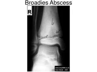
Calcaneus : Anterior process avulsion fracture • Pain post sprain • Easily missed on X-rays • High index of suspicion • Scrutinize X-rays • Bone tenderness always present • Rx : Open excision if problematic
Talus Fractures • Osteochondral • Shear / sagittal / coronal • Posterior process • Os trigonum • Posteromedial ( Cedell ) / posterolateral process • Lateral process
Lateral Talar process fx • “Snowboarder’s fracture ” • Diagnosis delayed & associated with ankle sprains • Need a high degree of suspicion
Treatment : lateral process # • Acute - nondisplaced: cast treatment NWB • Acute - displaced: ORIF or excise • Late: excise or ORIF based on size ( usually chronic subfibular pain ) • Excise open or arthroscopic
Lateral talar process avulsion • Rx : excision
Periarticular ( avulsion ) Fractures Sports Trauma Summary: • Common cause of Chronic dysfunction / pain • Ankle arthroscopy is an excellent procedure for evaluation and treatment • minimal morbidity with careful technique • Excision is the common treatment , unless fixation warranted
Ankle Arthroscopy Acute AnkleFractures: • Advantages • avoids extensive exposure • improves visualization of articular surface • maintains existing blood supply Disadvantages • time consuming • technically more challenging • swelling of soft tissues
Ankle Arthroscopy Acute AnkleFractures: • Indications • Mild to moderate pilon fractures/ impaction • To ensure articular surface reduction • Remove loose fragments/ hematoma/ chondral injury
Post op 1 year post op
Literature Review • Hintermann B, Regazzoni P, Lampert C, Stutz G, Gachter A. • Bone Joint Surg Br. 2000 Apr;82(3):345-51. • Arthroscopic findings in acute fractures of the ankle. • Prospective study • Ankle # in 288 consecutive patients (148 men and 140 women) • AO-Danis-Weber , 14 type-A,198 type B and 76 type C. • Chondral lesions in 228 ankles (79.2%), the talus (69.4%) ;distal tibia (45.8%), the fibula (45.1%), medial malleolus (41.3%). • worse in patients under 30 years and in those over 60 years of age. • The frequency and severity of the lesions increased from type-B to type-C fractures (p < 0.05).
Literature Review : ARIF Ankle # • Ono A, Nishikawa S, Nagao A, Irie T, Sasaki M, Kouno T. • Arthroscopy. 2004 Jul;20(6):627-31. • Arthroscopically assisted treatment of ankle fractures: arthroscopic findings and surgical outcomes. • 105 patients (105 joints) ; malleolar fractures • Cartilaginous damage was noted in 21 patients • distal tibiofibular joint diastasis + fixation in 8 patients. • good result in 100 cases and a fair outcome in 5 • (no control group).
Use of Ankle Arthroscopy with Fractures Summary • Useful adjunct in diagnosis and treatment • Biologic exposure • Needs further experience and investigation