Download
1 / 57
570 likes | 672 Views
Chapter 2 Epithelial tissue. 1.General feature: 1) contain more cells and less extracellular ground substance 2) Polarisaton : ---free outer surface: face the surface of the body or the lumen of an organ or gland ---basal surface: face underlying connective tissue

E N D
1.General feature: 1) contain more cells and less extracellular ground substance 2) Polarisaton: ---free outer surface: face the surface of the body or the lumen of an organ or gland ---basal surface: face underlying connective tissue 3) Avascularity, but innervation: ---no blood vessels ---rich in nerve terminals 4) Cellular Layer + Basement Membrane 5) Functions: protection secretion absorption sensory reception
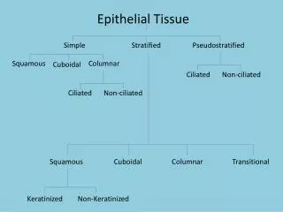
2.Classification of Epithelium 1)Covering epithelium: the epithelium which cover body surface or line the inner surface of body cavities, tubes and sac. 2)Glandular epithelium: the epithelium which main function is secretion. 3)Sensory epithelium: the epithelium which has special sensory function. * Myoepithelial cell
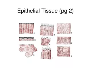
3. Classification of covering epithelium: According to the number of layer and shape of cells Simple epi.: ---simple squamous epi. ---simple cuboidal epi. ---simple columnar epi. ---pseudostratified ciliated columnar epi. Stratified epi.:---stratified squamous epi. ---stratified columnar epi. ---transitional epi.
Cell Layers Simple Stratified
1)simple squamous epi: ---structural feature: /one layer flattened cells, cell border are interdigitate closely /with flattened ellipsoid nucleus
---Distribution: • mesothelium: the simple squamous epi. which line the inner surface of body cavities such as thoracic, abdominal cavities and pericardiac . • endothelium: the simple squamous epi. which line the inner surface of cardiovascular and lymph vessels. • other place: alveoli, parietal layers of renal capsule. ---function: a) transport of materials b) facilitates movement of viscera
2)simple cuboidal epi.: ---structural feature: • one layer of cells, with same height and width , hexagonal in shape. • spherical centrally-located nucleus
---distribution: /the renal tubule /thyroid /the some ducts of glands ---function: covering and secretion renal tubule thyroid
3)simple columnar epi.: ---structural features: • one layer of columnar cells, with basally located ovoid nucleus
---distribution: gastrointestinal tract gall bladder uterus ---function: secretion and absorption goblet cell: scattered, secreting granules-mucus goblet cell simple columnar epi
four types of cells 4)pseudostratified ciliated columnar epi.: ---Structural feature: 1, Four types of cells columnar cell (ciliated); goblet cell fusiform cell; basal cell: pyramid-shaped 2, Every cell locate on basement membrane: Simple epi.
---distribution: inner surface of large duct of respiratory passages trachea bronchi nasal The epithelium of trachea
5)stratified squamous epi.: ---structural features: • deepest (basal) cells: one layer of cuboidal cells • the cells in intermediate regions: several layers of polygonal –shaped cells • to the surface: more and more flattened cells
---distributon: • non-keratinised: mouth, pharynx, oesophagus, urethra and vagina • keratinised: the surface of body, make up the skin karatinised non-karatinised
6)transitional epithelium: • Flexible qualities ---ncluding the number of layers and shape of cells
in the contractedbladder in the distendedbladder
4. Epithelial specializations • Sides of cells: • --- Apical (free) • --- Basal • --- Lateral
①microvilli: ---definition: delicate finger-liked projections of cell-membrane and cytoplasm protruding from the free surface
---structure: 0.1um in diameter, with different longth. surface: cell membrane with cell coat core: longitudinal microfilament-actin filament fixed on terminal web terminal web: made up of transverse-arranged filament at the apical side of cells
---function: increase the surface areas, thus aid in the processes of secretion and absorption. ---distribution: /striated border: intestinal epi. cell /brush border: proximal renal tubule Striated border
②cell coat: ---definition: a thick layer of extracellular glycoprotein ---function: adherence, supporting, protection, exchange of material and recognize
③ cilia: ---definition: elongated, mobile projections of cell membrane and cytoplasm protruding from free surface
---structure: • 5-10um long, 300-500nm in diameter • surface: cell membrane • core: microtubules, 9X2+2 • basal body: centrioles-connected with microtubules of cilia
---function: beat in a rhythmical manner and produce a forward-moving wave ---distribution: epithelial cells of respiratory tract respiratory tract
---intercellular connection of adjacent cells: • non-special manner: the minute space and adherent molecules (glycoproteins, proteoglycans, cadherin) • Special manner: junctional structures
①Tight junction (zonula occludens): ---structure: • apical part • point-liked fused between adjacent cells • arranged in 2-4 thread-liked structures • form anastomosing network ---function: seal the space between cells
② intermediate junction (zonula adherens): ---structure: • below the tight junction • a gap of 15-20nm in width with medium electron-density filament material • plaque of electron-dense materials, with attached microfilament-make up of terminal web ---function: /adherens /keep the cell shape /transfer cell contract force
③desmosome (macula adherens): ---structure: • plate or spot-shaped • a gap of 20-30 nm, with low electron-density filaments interdigitate • attachment plaque • Many tonofilaments are inserted into attachment plaque, each filament make a hairpin loop and then passes back into the cytoplasm • ---function: firmly connection
④gap junction (communicating junction): ---structure: • the smallest gap of 2-3 nm • connexons: -consist of protein -7~9nm in diameter -composed of 6-subunits of proteins- connexin -2nm channel: electron-lucid central channel ---function: provide a ions and small molecules pathway between cells
junctional complex: at least two types of junctional structures get together.
①basement membrane: ---definition: a sheet of membrane-liked amorphous material interposed between epithelium cells and underlying CT. ---structure: • HE: pink colour, hard to see • PAS +
EM: two layers --basal lamina: 20-300 nm, electron-dense, thread-liked and amorphous ground substance, produced by epithelium Cell --reticular lamina: RT+ground substance, produced by CT
---function: • support, connection, fixation • semi-permeable membrane • induce the movement, proliferation and differentiation of epithelium cell
② plasma membrane infolding (basal longitudinal striation): ---definition: the infolding of cell-membrane with many mitochondria at the basal surface of epithelium cell
---function: • increase the basal surface areas • facilitate the passage of water and ions ---distribution: mainly in proximal and distal renal tubule.
③hemidesmosomes ---is half of desmosome.
5. Glandular epithelium and gland (study yourself ) • glandular epithelium: are specialized for secretion • gland: organs composed mainly of glandular epiithelium
1) Classification: exocrine gland: discharge the secretion through a duct system endocrine gland: release the secretion directly into blood steam
Simple gland compound alveolar gland compound tubulo-alveolar gland 2) structure of exocrine gland: