Download
1 / 14
160 likes | 396 Views
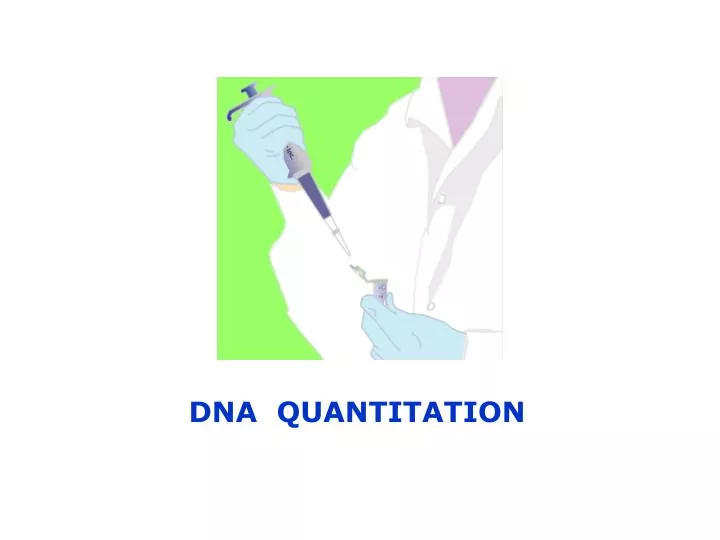
DNA QUANTITATION. 2 methods for DNA Quantitation. I. Agarose Gel Electrophoresis. II. Spectrophotometer. Gel Electrophoresis. - Electrophoresis is a technique used for separation of charged molecules.

E N D
2 methods for DNA Quantitation I. Agarose Gel Electrophoresis II. Spectrophotometer
Gel Electrophoresis - Electrophoresis is a technique used for separation of charged molecules. - DNA is a negatively charged molecule, and is moved by electric current through a matrix of agarose.
Agarose Gel extracted from seaweed linear polymer structure
Mobility of DNA Molecules DNA has a negative charge because of the phosphate backbone It migrates in an electric field from negative charge to positive charged cathode
Circular form of DNA Linear DNA Supercoil DNA Mobility of DNA Molecules Shape & Size I. Conformation of DNA Supercoil DNA migrate faster than circular DNA and linear DNA migrate slowly
Mobility of DNA Molecules II. Length of DNA fragment
Mobility of DNA Molecules Gel Concentration Higher concentrations of agarose facilitate separation of small DNA Low agarose concentrations allow resolution of larger DNA
Anode (+) Cathode (-) As the voltage applied to a gel is in creased, the DNA fragment migrate faster than low voltage. Voltage Applied
Electrophoresis Buffer TAE (Tris-acetate-EDTA) TBE (Tris-borate-EDTA) Loading Buffer 6x Loading Dye 0.25% Bromophenol blue – BB— (or tiny amount on the spatula tip) 0.25% Xylene cyanol FF –XC— (or same as BB) 15% Ficoll (Type 400)
Visualization of DNA Fragments Ethidium Bromide
Std.100 ng std. 300 ng std. 500 ng std. 1000 ng KDML105-1 KDML105-2 KDML105-3 IR62266-1 IR62266-2 IR62266-3 BT-1 BT-2 BT-3 IR62266(6) Standard of DNA concentration Samples - +