Download
1 / 47
550 likes | 976 Views
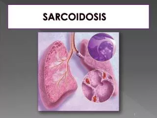
Azin Alizadehasl , MD. Echocardiography in sarcoidosis. Sarcoidosis is a systemic inflammatory disease of unknown etiology, characterized by non- caseating granulomas . It mainly affects people in the 3th and 4th decades of life, but may also be found in children or elderly subjects.

E N D
AzinAlizadehasl, MD Echocardiography in sarcoidosis
Sarcoidosisis a systemic inflammatory disease of unknown etiology, characterized by non-caseatinggranulomas. It mainly affects people in the 3th and 4th decades of life, but may also be found in children or elderly subjects.
Braunwald heart disease: Sarcoidosis: RWMA Thining : basal of posterior and lateral walls Posterobasal aneurysm Restrictive morphology PAH and right side failure
Feigenbaum’s Echo: The 2D echo features of myocardial involvement occur in less than 20% of sarcoid patients. Heart is involved in as many as 50% of advanced cases. LVE+RWMA(base and mid levels).
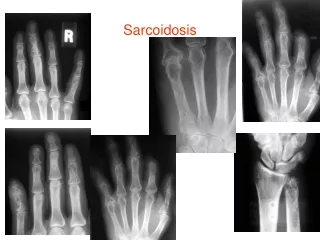
Otto: • Cardiac involvement include: pericardium, conducting system myocardiom ( microscopic focal infiltration or larger nodules). • Basal posterior and lateral walls and septum. • MR is not uncommom.
RWMA is often in a location inconsistent with unusual coronary anatomy. Disseminated sarcoidosis: DCM There are no specific echo findings in cardiac sarcoidosis.
Heart involvement has been reported in as many as 58% of pts and may be responsible for up to 85% of deaths due to sarcoidosis.
Autopsy studies and various types of examination reveal a high prevalence of heart involvement, • Clinical manifestations and symptoms are rare (about 5% of cases).
They include advanced AVB, malignant ventricular arrhythmia, MR due to papillary muscle dysfunction, pericarditis, CHF and SCD.
No single test or investigation can detect specific abnormalities enabling diagnosis. • Various methods including CMR, thallium-201 and gallium have been suggested as valuable diagnostic tools.
Various forms of echocardiography have been tried and features such as septal thinning, LV regional systolic dysfunction, • pericardial effusion and commonly LV diastolic dysfunction determined, but these abnormalities only seem to be present in advanced heart involvement.
Pts and controls had similar LV diameters, wall thicknesses, LA size, eFS and EF.
Doppler echo evidence of LV diastolic dysfunction was detected in 33 (55%) pts.
mFS(mid wall fractional shortening) was determined • using a prolate ellipsoidal model of LV geometry; • LV wall is divided into inner and outer shells with equal thickness at diastole.
Sarcoid pts had lower mFS than controls (15.8 ± 2.4% vs. 18.8 ± 2.5%, p=0.001.
Pts with a longer history of the illness had lower mFS (16.6 ± 2.8% vs. 15.1 ± 1.9%, p = 0.024).
Follow-up of our pts will show whether a lower mFS may have prognostic value in these pts.
LV systolic dysfunction and RWMA in pts with severe heart involvement. when the disease only involved the conduction system echocardiography was substantially normal.
Pts with diastolic dysfunction may have had granulomas in the myocardial interstitium, which could stiffen the myocardium and damage ventricular Relaxation.
TDI demonstrated impaired longitudinal strain and strain rate of several lateral LV segments. • 2D echo showed loss of wall thickness and hypokinesia of these segments.
PAH • The frequency of PH is ≈20%. • The presence of PH contributed to poor outcomes in pts with sarcoidosis.
Case report(JACC 2008): A 59-year-old woman with sarcoidosis was admitted to hospital for assessment of AVB3. Echo showed RV dilation and dyskinesis of the apical free wall, whereas LV study showed normokinesis, mimicking RV dysplasia.
Granulomas are most frequently located in the LV free wall (96%), followed by the interventricular septum (73%); whereas, the atrial wall is rarely affected (right, 11%; left, 7%).
40% of CS pts had increased echoes(local scaring) and hypokinesis of the proximal part of interventricle septum.