Download
1 / 19
350 likes | 1.4k Views
Chronic Obstructive Pulmonary Disease. Margaret Saunders RN, BSN, CCRN Liberty University. Objectives. Provide a brief introduction of Chronic Obstructive Pulmonary Disease(COPD) and it’s relationship to chronic bronchitis and emphysema. Discuss the incidence and prevalence of COPD.

E N D
Chronic Obstructive Pulmonary Disease Margaret Saunders RN, BSN, CCRN Liberty University
Objectives • Provide a brief introduction of Chronic Obstructive Pulmonary Disease(COPD) and it’s relationship to chronic bronchitis and emphysema. • Discuss the incidence and prevalence of COPD. • Review the pathophysiology of bronchitis and emphysema as it relates to the adult client. • Discuss diagnosis and management of COPD. • Provide patient education as it relates to prevention, pharmacological, emotional and end-of-life care.
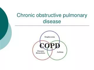
Introduction • Chronic obstructive pulmonary disease (COPD) is a respiratory disease of inflammation. It is used to describe a progressive and irreversible decrease in lung function(Higginson, 2010). This decline in lung function results in decreases airflow and obstruction. The term COPD can be used to describe the more specific terms, emphysema and chronic bronchitis. • The primary risk factor for COPD is smoking. Other risk factors consist of air pollution, second-hand smoke, occupational exposure to toxins, and frequent respiratory infections (Higginson, 2010). • At present time there is no known cure for COPD, however with treatment progression of the disease can be controlledand quality of life improved(McCance, Huether, Brashers, & Rote, 2010).
Incidence & Prevalence • COPD is the most common lung disease in the United States. The current data estimates 13.1 million adults aged 18 and over have been diagnosis with COPD. However, there may be as many as 24 million adults with some evidence of impaired lung function, suggesting an under diagnosis of this disease(American Lung Association (ALA), 2011). • COPD is more prevalent among non-Hispanic white and Puerto Rican adults than those of non-Hispanic black and Mexican-Americans(Akinbami & Liu, 2011). • COPD is the third leading cause of death in America, claiming the lives of 124,477 Americans in 2007(American Lung Association (ALA), 2011; Han, 2011). • The total deaths from COPD is projected to increase by 30% in the next 10 years without any interventions to decrease the risk, particularly smoking(WHO, 2011).
Genetics & Genomics • COPD is a result of the combination of risk factors and host susceptibility. • Genetic susceptibilities have included mutations in TNF, surfactant, proteases, and anti-proteases(McCance et al., 2010). • There is a noted link between COPD and Alpha-1 antitrypsin; with a deficient of Alpha-1 antitrypsin has been shown to increase the likelihood of developing emphysema. (Higginson, 2010). • Research in COPD is certainly on the move in many different areas, but more than ever the coordination and integration of information is needed to provided a greater understanding of COPD(Han, 2011).
Risk Factors • COPD is primarily caused by smoking. • Additional risk for developing COPD includes: air pollutants, occupational exposure to toxins, and history of severe respiratory infections. • The development of COPD as an early age has been linked to the inherited mutation in the Alpha-1 antitrypsin gene.
Emphysema • Emphysema causes the walls between the alveoli to lose their ability to stretch and recoil. The air sacs become stiff and weakened and may break, creating irreversible “holes” in the tissues of the lower lungs. These holes between the small air sacs create larger air sacs, in which air can become trapped more easily. The lungs have more difficulty moving air in and out and the exchange of oxygen and carbon dioxide with the blood may be impaired (McCance et al., 2010).
Emphysema Pathophysiology • According to McCance Huether, Brashers, & Rote (2010), approximately 1-2% of emphysema cases are a result of an inherited lack of alpha-1 antitrypsin. Alpha-1 antitrypsin is a protein that protects the lungs by inhibiting neutrophil elastase(Higginson, 2010). Known as primary emphysema, because those with deficiency in Alpha-1 antitrypsin have an increased risk of developing emphysema, especially if those individuals smoke (McCance et al., 2010). • Secondary emphysema results from insults to the lungs from inhaled toxins, such as smoke or air pollution. The inhaled toxins led to inflammation of the epithelium, stimulating cytokines to release proteases and the inhibition of antiproteases in the lung tissue (McCance et al., 2010). As noted in Higginson(2010), this imbalance leads to the breakdown of elastin causing destruction of the alveoli wall, limiting gas exchange and reducing elastic recoil. As elastic recoil decreases air becomes trapped within the lungs. The trapped air causes hyper expansion of the chest and increase work to breath so many individuals will develop hypoventilation, hypercapnia, and hypoxia. With the progression of the hypoxia leading to pulmonary hypertension and eventually cor pulmonale(Higginson, 2010).
Clinical Manifestations • Emphysema “pink puffers” • Dyspnea on exertion that progresses to finally the client will have dyspnea at rest. • Little coughing and little if any sputum. • Client is often thin in appearance with a barrel chest. • Tachypnea with prolonged expiration and use of accessory muscles. • Client often sits leaning “tripod position” forward to increase lung capacity. • Pursed-lip breathing (McCance, Huether, Brashers, & Rote, 2010)
Chronic Bronchitis • Inflammation and eventual scarring of the lining of the bronchial tubes. This initiates the increase production of mucous leading to a reduction in bronchial tube inter-diameter decreasing the flow of air to and from the lungs (American Lung Association, 2011).
Chronic Bronchitis Pathophysiology • As defined by McCance et al(2010) chronic bronchitis is a hyper secretion of mucus and chronic productive cough that occurs for at least 3 months of the year for 2 consecutive years. • The inspired toxins(smoke) leads to a defensive increase in mucous production. This increase in mucous production results in an increase number of goblet epithelial cells and impaired ciliary function, resulting in mucous gland hyperplasia, increasing the risk for infection(Higginson, 2010). • The continued inflammation and recurrent infections results in the progression of the disease leading bronchospasm and permanent wide spread narrowing in the small airways, increasing airway resistance and fibrosis around bronchioles. (McCance et al., 2010).
Clinical Manifestations • Chronic Bronchitis “blue bloaters” • Decreased exercise tolerance, wheezing, and dyspnea. • Clients usually have a productive cough “smoker’s cough” • Decreased forced expiratory volume in one second (evidence of airway obstruction). • With the progress of the disease the forced volume capacity and forced expiratory volume continue to decrease, while the residual volume increases as with airway obstruction and air trapping. • Airway obstruction causes decreased alveolar ventilation and increased Paco2. • Cyanosis and polycythemia due to the pronounced hypoxemia. (McCance, Huether, Brashers, & Rote, 2010)
Diagnosis/Evaluation • A complete history and physical should be completed to assess for risk factors, respiratory changes, and overall general appearance. • Arterial blood gas to identify abnormal oxygenation and ventilation. Elevation of the PaCO2 and a reduced PaO2 level can be noted with COPD. • Pulmonary function tests indicate a marked decrease in forced expiratory volume suggesting obstruction in gas flow during expiration(McCance, Huether, Brashers, & Rote, 2010). • Chest radiograph demonstrates a flattened diaphragm and lung fields appear over distended. (McCance et al., 2010)
Treatment/Care • Goals of treatment consist of the following: relief of symptoms, slow the progression of the disease, improve overall health (exercise tolerance) and the prevention of complications and exacerbations (McCance, Huether, Brashers, & Rote, 2010). • Medications do not prevent the progression of COPD, but will reduce the extent of dyspnea a client experiences. • Anticholinergic Bronchodilator -relax airway muscles by blocking the acetylcholine. The most commonly short-acting anticholinergic prescribed is Atrovent, used as a meter-dosed inhaler. Spiriva is a long-acting anticholinergic that may beneficial given daily in combination with a beta2 adrenergic agonist (Katz, 2010). • Beta2 Adrenergic agonist -acts as a bronchodilator. The short-acting form of beta2 adrenergic agonist are used as rescue medication to provide immediate relief of dyspnea. Albuterol is the most commonly prescribed short-acting bronchodilator, taking effect in 5 to 15 minutes. Serevent is a long-acting beta2 agonist providing relief for at least 12 hours (Katz, 2010).
Treatment/Care cont… • Corticosteroids -are helpful to decrease airway inflammation, however the continued use of oral steroids can cause other problems(Cushing’s syndrome, glaucoma, hyperglycemia, and the inability to overcome infections) (Katz, 2010). Flovent, a commonly used inhaled steroid has been helpful in reducing airway obstruction. Such, inhaled corticosteroids are usually reserved for the client with severe COPD(Katz, 2010). • The phosphodiesterase inhibitor, theophylline, is used to dilate airways, relax smooth muscles, stimulate the respiratory of the brain and improve function of the respiratory muscles. Theophylline usually given orally and is the last bronchodilator to be added to the treatment regimen (Katz, 2010). • Oxygen therapy is used to improve oxygenation and ease the work of breathing. Oxygen along with smoking cessation are the only strategies proven to improve survival in the clients with COPD (Higginson, 2010). • Chest physical therapy in combination with postural drainage could provide a method to clear the increase secretions(Katz, 2010). • Surgery is a last resort for those with COPD (McCanceet al., 2010).
Patient Education • Education should be given in relation to disease process and medications to improve dyspnea. Pulmonary rehabilitation is suggested to help clients; with the aim to reduce symptoms and disability associated with COPD (Higginson, 2010;McCance et al., 2010). • Lifestyle change should include: smoking cessation, which includes second-hand smoke that causes increase irritation and inflammation of the lungs(Higginson, 2010). • Nutrition is discussed and a balance diet including protein and adequate fluid is encouraged. If dyspnea hinders eating, suggest small and frequent meals allowing time for rest during meal. • Vaccinations are recommended to decrease the likelihood of complications associated with COPD. • Exercise consisting of physical activity as tolerated by the client. Instruction and demonstration of breathing techniques (diaphragmatic breathing as well as pursed-lip breathing) to help reduce dyspnea (Katz, 2010). • Anxiety is often noted in COPD, due to disease progression and the increase work of breathing and dyspnea. Depression may be of concern to the client and should be addressed. Emotional support is given to the COPD client. Early discussion of palliative care will give options to the client regarding end-of-life considerations, easing the decisions for family members. Severe difficulty in breathing is uncomfortable and upsetting to the client and family members. “Near the end of a COPD client’s life, dyspnea must be eased” (Katz, 2010).
References • American Lung Association (ALA). (2011). Chronic obstruction pulmonary disease (COPD) fact sheet. Retrieved from http://www.lungusa.org/lung-disease/copd/resources/facts-figures/COPD-Fact-Sheet.html • Akinbami, L. J., & Liu, X. (2011). Chronic obstructive pulmonary disease among adults aged 18 and over in the United States, 1998-2009. NCHS data brief no.63. Hyattsville, MD: National Center for Health Statistics. Retrieved from http://www.cdc.gov/nchs/data/databriefs/db63.htm • American Lung Association. (2011). Trends in COPD (chronic bronchitis and emphysema): Morbidity and mortality. Retrieved October 4, 2012, from http://www.lung.org/finding-cures/our-research/trend-reports/copd-trend-report.pdf • Han, M. K. (2011). Update in chronic obstructive pulmonary disease in 2010. American Journal of Respiratory and Critical Care Medicine, 183, 1311-1315. doi:10.1164/rccm.201102-0280UP
References cont… • Higginson, R. (2010). COPD: pathophysiology and treatment. Nurse Prescribing, 8(3), 102-110. • Katz, M. J. (2010). Chronic obstructive pulmonary disease (COPD). Retrieved 2012, from Wild Iris Medical Education: http://www.wildirismedicaleducation.com/courses/297/index_nceu.html • McCance, K., Huether, S., Brashers, V. L., & Rote, N. S. (2010). Pathophysiology: The biological basis for disease in adults in children (6th ed.). St. Louis, MO: Mosby. • WHO. (2011, November). Chronic obstructive pulmonary disease (COPD) Fact Sheet N315. Retrieved October 02, 2012, from World Health Organization: http://www.who.int/mediacentre/factsheets/fs315/en/index.html