Download
1 / 33
460 likes | 998 Views
Chronic obstructive pulmonary disease. Chronic obstructive pulmonary disease (COPD). Permanent reduction in airflow in the lung Caused by smoking, air pollution, dust, lack of alpha 1 -antitripsien. COPD Patho physiology. Loss in elasticity due to changes in

E N D
Chronic obstructive pulmonary disease (COPD) Permanent reduction in airflow in the lung Caused by smoking, air pollution, dust, lack of alpha1-antitripsien
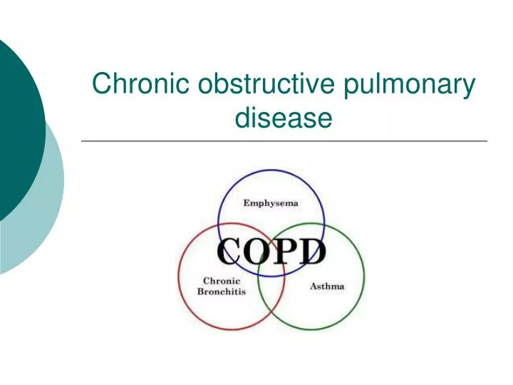
COPDPatho physiology Loss in elasticity due to changes in collagen and elastin on alveolar level Narrowing of airways
COPDChronic bronchitis Productive cough for more than 3 months of 2 consecutive years (other conditions excluded)
Chronic bronchitisPathology ↑ mucous production Hypertrophy of mucous glands Thickening of the airway ↑ number of goblet cells Thus narrowing of the lumen of the airways and airway obstruction. Infection caused by accumulated secretions.
COPDEmphysema Permanent enlargement in the normal size of the air spaces distal to the terminal bronchioles due to destruction of alveolar tissue.
EmphysemaPathology Lack of alpha1-antitripsien causes uncontrolled breakdown of collagen and elastin, damaging the alveolar framework
EmphysemaClassification “Blue-bloater” Moderately severe airflow impairment Stimulus for breathing ↓ PO2
EmphysemaClassification “Pink puffer” Little sputum production, dyspnoea gr.IV Right heart failure and peripheral oedema
Emphysema and Chronic bronchitisClinical signs Use of accessory muscles Drawing in of supraclavicular fossae and intercostal space ↓ chest expansion ↓ lung sounds (breath sounds) Dyspnoea with or without productive cough
Emphysema and Chronic bronchitisX-rays Hyperinflation Flattened diaphragms Lengthening of heart shadow Prominent hilar vessels
EmphysemaLung functions ↓ FEV1 ↓ forced vital capacity ↓ peak flow ↑ total lung capacity and residual volume
EmphysemaCourse of disease Airflow impairment develops over long time Productive smoker’s cough Acute bronchitis Cannot go to work – severe bronchitis Attacks occur repeatedly – lose jobs
EmphysemaComplications Cor pulmonale – pulmonary hypertension causes right ventricular failure Bullae – alveolar walls burst and form large air-filled spaces with thin walls
COPD rehabilitationDyspnoea Overactivity of accessory muscles inhitis diaphragm Patient must be taught to breathe with lower part of his chest
COPD rehabilitationDyspnoea Relaxation positions and breathing control “Pursed lip breathing”
“Pursed lip breathing” • Maintains airway pressure in lungs, prevents airways from collapsing • ↑ airflow
COPD rehabilitation Bronchodilators Relieves bronchospasm Anti-cholinergic drugs (atrovent) and not B2-stimulants If stimulus for breathing is ↓PO2 – do not nebulise with 100% O2
COPD rehabilitation Improve exercise tolerance Improve physical activity to highest functional level Improve quality of life 6 minute walking test Exercise programme
COPD rehabilitation Remove secretions Nebulise with mucoliticum Percuss, shake and vibrate Precaution – patients on korticosteroids develop osteoperosis. Shaking and vibrating can cause rib fracture. “Huffing”
“Huffing” • Forced expiratory technique • Just as effective as coughing, less effort • Medium-sized breath, mouth and glottis open, force air out using chest wall and abdominal muscles.
References • Pryor, J.A. and Prasad, S.A. 2009. Physiotherapy for respiratory and cardiac problems. Adult and paediatrics. Edinburgh: Churchill Livingstone • FTB 309 Dictate • Images courtesy of Google search engine