Download
1 / 38
390 likes | 692 Views
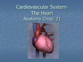
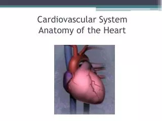
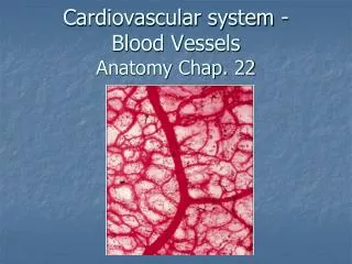
Anatomy of the Cardiovascular System. Chapter 18. Heart. Location: mediastinum, just behind the body of the sternum, between second-sixth ribs. 2/3 of mass is left of the midline of the body Posteriorly rests against the fifth to eighth thoracic vertebrae

E N D
Anatomy of the Cardiovascular System Chapter 18
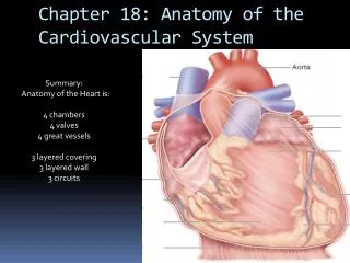
Heart • Location: • mediastinum, just behind the body of the sternum, between second-sixth ribs. 2/3 of mass is left of the midline of the body • Posteriorly rests against the fifth to eighth thoracic vertebrae • Because lies between boney structures, Cardiopulmonary Resuscitation (CPR) may be done to save life
Size and Shape • Shape of the adult heart determined by the shape of the chest • Tall and thin: heart is elongated • Short and stocky: heart is wide (transverse) • Average build: heart shape intermediate • Size of the adult heart determined by sex • Male heart larger than female • Adult male (avg) is ~310 g • Adult female (avg) is ~224g
Coverings of the heart • Pericardium • A loose fitting inextensible sac • Two parts • Fibrous pericardium • Tough loose-fitting, and inelastic sac around the heart • Attaches to the large blood vessels emerging from the top of the heart but not to the heart • Serous pericardium • Parietal layer: lining inside of the fibrous pericardium • Visceral layer (epicardium): adhering to the outside of heart; between visceral and parietal layers is a space (pericardial space) which contains 10-15 ml of fluid (pericardial fluid)
Functions: • Fibrous pericardial sac provides protection against friction • Serous pericardium produces the lubricant required to reduce friction
Structure of the Heart • Wall • Epicardium: outer layer (“on the heart”) • Is the visceral layer of the serous pericardium • Myocardium: middle layer • Is a thick, contractile of specially constructed cardiac muscle cells • Encircling muscle structure that compresses the heart cavities, and the blood, with great force and without fatigue (autorythmic)
Endocardium: interior of the myocardial wall • Made of a membranous tissue that that lines the heart and blood vessels. • Covers beamlike projections of mycardial tissue called trabeculae • Folded layers of pockets of endocardium make the majority of the valves used to regulate the flow of blood
Chambers of the heart • Atria • Two superior chambers of the heart • The receiving chambers of blood from veins • Thin walled because only move blood a short distance (into the lower chambers) • An earlike flab protruding from each atrium is called an auricle (ear-like)
Ventricles • Two lower chambers of the heart • Primary pumping chambers—pump blood into the arteries to be carried throughout the body • Thick walled, left thicker than right • Left must pump blood out to the rest of the body • Right must only pump to lungs
Valves of the heart • Atrioventricular valves • Right side • Made of three cusps (flaps) of endocardium • Free edge anchored to papillary muscles of right ventricle with chordae tendineae • More commonly called the tricuspid valve • Left side • Made of two cusps (flaps) of endocardium • Free edge anchored to papillary muscles of left ventricle with chordae tendineae • More commonly called the bicuspid valve • Atrioventricular valves allow blood to flow from the atria into the ventricles but prevents it from flowing back up into the atria from the ventricles
Semilunar valves • Half moon-shaped flaps growing out from the lining of the4 pulmonary artery and aorta • Pulmonary semilunar valve: entrance of the pulmonary artery • Aortic semilunar valve: valve at the entrance of the aorta • Closure of the semilunar valves prevents backflow of blood into the ventricles
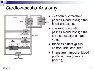
Flow of blood through the heart • Blood enters the heart from the body through the right atrium. • Blood then flows to the right ventricle through the tricuspid valve. • Right ventricle contracts and pushes blood through the pulmonary semilunar valve into the first portion of the pulmonary artery. • Artery branches to form right and left pulmonary arteries—go to right or left lung. • Gas exchange takes place, blood flows to through the right and left pulomonary veins to the left artrium. • Left atrium contracts and enters into the left atrium through the bicuspid valve (mitral) • Left ventricle contracts sending blood through the aortic semilunar valve into the aorta • Aorta branches to supply all areas of the body with oxygenated blood • Cell waste (deoxygenated blood) enters into veins • All veins from head and neck region empty into the superior vena cava; all veins from below the neck enter into the inferior vena cava • Superior vena cava and inferior vena cava empty into the right atrium
3D reconstruction of the heart as viewed from the apex towards the valves, image flipped 180° relative to illustration above. Pulmonary valve not visible, leaflets of the tricuspid and aortic valves only partly visible. To the left two images in 2D from the same dataset, showing tricuspid and mitral valves (above) and aortal and mitral valve (below).
Heart Blood Supply • Coronary arteries • Right and left • Lie between the aortic semilunar valve • Both ventricles receive blood from these • Each atrium receives blood only form a small branch of the corresponding coronary artery • Most abundant supply goes to the myocardium of the left ventricle • Right coronary artery is dominant in half of all hearts, left only dominant in about 20% • Heart has only a few anastomosis (connections from the proximal part of an artery to a more distal part of itself or another artery)
Few anastomosis does not allow oxygenated blood to reach the heart if main artery becomes blocked • Myocardial infarction (death of ischemic (blood deprived) cardiac cells) ***Bypass surgery created to prevent this*** Video links
Cardiac Veins • Deoxygenated blood travels through capillaries from the myocardium to the cardiac veins • Veins empty into the right atrium
Conduction System of the Heart • Sinoatrial Node • SA Node or pacemaker • In right atrial wall near opening to superior vena cava • Atrioventricular Node • AV Node or Node of Tawara • In right atrium along the lower part of the interatrial septum • Atrioventricular Bundle • AV Bundle or bundle of His • Originate in the AV Node and extend by two sides of interventricular septum • Purkinje fibers • Continuation of the AV Bundle • Extend out lateral walls of ventricles and papillary muscles
Conduction structures consists of cardiac muscles modified enough in structure to differ from ordinary cardiac muscle function • Permit only the generation or rapid conduction of an action potential through the heart • Set the rhythm of the heart
Heart Nerve Supply • Sympathetic Fibers (accelerator nerves) • Contained in middle, superior, and inferior cardiac nerves • Parasympathetic Fibers (Vagus fibers, inhibitory or depressor nerves)) • Located close to the arch of the aorta **Allow for the heart to be controlled by the autonomic nervous system**
Blood Vessels • Artery (arterioles) • Vessel that carries blood away from the heart • All but the pulmonary artery carry oxygenated blood • Vein (venules) • Vessel that carries blood toward the heart • All but the pulmonary vein carry deoxygenated blood • Capillaries • Vessel that carries blood from arteries to veins • Only one cell thick, carry oxygenated blood to cells from artery; carry cell waste to veins • Some areas have spaces that take place of capillaries, these are called sinusoids (liver)
Blood Vessel Structure • Outer Layer (tunica adventitia) • “coat that comes first” • Strong, flexible fibrous connective tissue that hold vessels open and prevents tearing of vessel wall • Thinnest layer in arteries, thickest layer in veins • Middle Layer (tunica media) • “Middle coat” • Smooth muscle with a layer of elastic connective tissue • Permit changes in blood vessel diameter • Thicker in arteries than in veins
Inner layer (tunica intima) • “innermost coat” • Made of endothelium that is continuous with the endothelium of heart lining • In arteries is completely smooth; in veins helps to form semilunar valves that maintain one directional blood flow • Capillaries have only one thin coat of endothelium with fenestrations (windows)
Learn the names of the major arteries and veins in the body---the worksheets you label
Fetal Circulation • Differs due to fact oxygen and nutrient exchange from Maternal blood • Grow two umbilical arteries, an umbilical vein, and the ductus venosus • Placenta remember functions as lungs and digestive organs to interchange gases, foods, and wastes • No maternal-fetal blood exchange occurs because each flows through own capillaries
Read page 581 • #1-6 describes fetal circulation
Disease of Cardiovascular System • Disease of pericardium • Pericarditis (inflamation of pericardium) • Caused by trauma, bacterial or viral infection, tumors • Causes layers of visceral and parietal layers of the serous pericardium to rub, causes severe chest pain • Fluid, pus, or blood may accumulate in the space between the two pericardial layers and impair pumping of the heart (pericardial effusion)—may lead to compression of the heart nown as cardiac tamponade • Symptoms: pain that increases with breathing or coughing, a rubbing sound heard, difficulty breathing, reslessness, and accumulation of pericardial fluid • Treatment: pericardiocentesis, antibiotics, non-steroidal, anti-inflammatory drugs (asprin)
Disease of valves • Rheumatic heart disease • Delayed inflammatory response to streptococcal infection • Cardiac valves and other tissues in the body may become inflamed (rheumatic fever) • If severe, may result in stenosis (valves become narrower than normal causing the slow down of blood flow in heart chamber)
Mitral valve prolapse (MVP) • Genetic basis causes defect in bicuspid (mitral valve), may also be caused by diseases like rheumatic fever • Flaps extend back and into the left atrium, causing incompetence (leaking) of the valve • Occurs in 1 out of every 20 people, usually asymptomatic • May suffer chest pain and fatigue
Aortic regurgitation • Blood ejects forward into the aorta, but also regurgitates back into the left ventricle due to defective aortic semilunar valve • Ventricle becomes swollen due to increased volume of blood, tries to compensate by beating harder • Causes stress to heart and may lead to myocardial ischemia (oxygen deprived heart) ***replacement of defective valves is called valvuloplasty***
Disease of myocardium • Coronary artery disease (CAD) • Many causes, all cause reduction of blood flow to the mycardial tissue • Blockage of blood flow causes heart cells to be deprived of oxygen • Cells die or become damaged causing myocardial infarction (MI)—heart attack • Angina pectoris—severe chest pain
Atherosclerosis • Hardening of the artieries • Lipids and other substances build up inside wall of vessels, calcify---causes wall to become hard and brittle • Causes: smoking, high fat and cholesterol diets, hypertension (high blood pressure), genetics
Congestive heart failure (CHF) • Left side of heart (ventricle) is inable to pump blood effectively • Decreases blood flow to body, body inturn retains fluids • Causes pulmonary edema that in turn causes right heart failure
Disorders of blood vessels • Arteries • Arterioscleosis (hardening of the arteries) • Ischemia occurs causing death to tissue (necrosis) • Large area of tissue death called gangrene • Causes: age, diabetes, high fat and cholesterol diets, hypertension, and smoking • Treatment: vasodialators, angioplasty, stents, clearing with lasers, drills, etc., bypass • Aneurysm • Section of artery becomes unusually wide • Promote blood clots (thrombi) • Thrombus may cause embolisms (blockage) • May burst causing severe hemorrhaging • Aneurysm in brain may cause stroke (cerebrovascular accident CVA)
Disorders of veins • Varicose veins • Enlarged veins, cause blood to pool rather than continue to heart • Commonly occur in superficial veins, often due to standing for long periods • Hemorrhoids (Piles) • Varicose veins in the anal canal • Straining during defecation, pregnancy common causes
Phlebitis • Vein inflammation • Thrombophlebitis is acute phlebitis caused by a clot • May break free causing embolism