Download
1 / 32
360 likes | 1.39k Views
HYMENOLEPIS DIMINUTA ECHINOCOCCUS GRANULOSUS ECHINOCOCCUS MULTILOCULARIS By S ree keerthi. HYMENOLEPIS DIMINUTA. COMMON NAME: rat tapeworm. Parasite of rat,mouse & man . HABITAT

E N D
HYMENOLEPIS DIMINUTA • ECHINOCOCCUS GRANULOSUS • ECHINOCOCCUS MULTILOCULARIS By Sreekeerthi
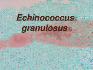
HYMENOLEPIS DIMINUTA COMMON NAME:rat tapeworm. • Parasite of rat,mouse & man. HABITAT • Adult worm resides in distal portion of ileum.
MORPHOLOGY • Worm measures 20 to 60cms in length • Larger than Hymenolepis nana • Scolex-4 suckers,retractilerostellum but has no hooklets. • Proglottids-800 to 1000.,mature proglottids contain ovary(centre),testis on either side. • Egg:infective to man,polar filaments absent
PATHOGENESIS • Rodents become infected by ingestion of arthropod hosts. • Man acquires infection accidentally by ingestion of infected fleas containing cysticercoid larvae.
CLINICAL MANIFESTATIONS • Most common in children. • Usually asymptomatic but noted symptoms are mild diarrhoea,abdominal pain & vague GIT manifestations.
LAB DIAGNOSIS MICROSCOPY -demonstration of eggs in faeces
TREATMENT • Praziquantel • Niclosamide. PROPHYLAXIS • Personal hygeine • Rodent control.
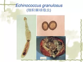
ECHINOCOCCUS GRANULOSUS COMMON NAMES • Hydatid tapeworm or dog tape worm. HABITAT • Adult worms are found attached to wall of intestinal mucosa of dogs $ wild canines • Larval stage found in humans $ herbivorous animals
HOOKLETS SCOLEX MORPHOLOGY M SUCKERS PROGLOTTIDS
PATHOGENESIS • Depending on modes of development cystic echinococcosis is of two types 1.primary cystic echinococcosis: -occurs after peroral infection with E.granulosus eggs that gives rise to hydatid cyst in different parts of the body. -most commonly these are found in liver and lung 2.secondary cystic echinococcosis: -occurs by rupture of primary hydatid cyst by trauama
Cont…………… -in this condition protoscolices are carried by blood circulation to different sites which develop into secondary hydatid cyst. The growing cyst surrounded by three layers : 1.pericyst containing fibrous tissue. 2.middle layer containing fibroblasts,eosinophils& blood vessels. 3.inner layer containing radially arranged gaint cells &eosinophils.
CLINICAL MANIFESTATIONS 1.Liver cysts :hydatidcyst in liver causes chronic abdominal discomfort,occasioally palpable or visible abdominal mass. • If cyst becomes infected with bacteria, it resembles an abscess • If cyst ruptures either spontaneously/trauma/surgery.There will be serious allergic reactions including skin rash,anaphylactic shock or death
2.lung cysts • Cysts are asymptomatic until they become large enough to cause cough,shortness of breath,chest pain. • Cyst rupture may lead to expectoration of hydatid fluid or membranes followed by the infection &lung abscess. • If rupture occur into the lung,it cause pneumothorax,empyema,allergic reactions & even anaphylactic shock
3.other sites • Spleen • Kidney • CNS • Bones • Heart • Muscles • FGT • Eyes
LAB DIAGNOSIS 1. CASONI skin test • It is immediate hypersensitivity skin test • Ag used is sterile hydatid fluid drawn from unilocularhydatid cysts from sheep , pig , cattle , man
Test arm Control arm Equal amt of sterile normal saline injected Control fades immediatly • 0.2 ml of antigen injected ID • In postive case develops large wheal, measuring 5cm or more in dia with multiple pseudopodia with in 30 mins
2. DLC may reveal eosinophilia20 to 25% 3. Serological test ELISA RIA CFT IHA LAT 4. Examination of cyst fluid 5. Histological examination 6.Radio diagnosis
Treatment • Surgical removal of hydatid cyst • Praziquantel & albendazole Prophylaxis 1. Strict personal hygiene 2. Reduction of stray dog population 3. Dogs should not b allowed to eat d carcase of slaughted animals
ECHINOCOCCUS MULTILOCULARIS MORPHOLOGY • Smaller than echinococcusgranulosus • 1.2-3.7mm • Eggs are typical Taenia like
PATHOGENESIS • Alveolar echinococcosis/Alveolar or multilocularHydatid cyst. • ORGAN-Liver. • Destruction of liver parenchyma-Hepatic failure. • Lesions spread to surrounding tissues $ body sites via Lymphatic/Haematogenous routes. • Central necrosis $ cavitation occurs $ cavity contains little or no fluid.
LAB DIAGNOSIS • BIOPSY of affected organ,identification depends on recognition of germinal & laminated membranes. • Radiodiagnosis:CTScan,Ultra sound. • Immunological Tests
TREATMENT • Surgical removal of Hydatid cyst • Praziquantel & Albendazole. PROPHYLAXIS • Strict personal hygiene. • Reduction of stray dog population.