Download
1 / 46
470 likes | 744 Views
Chromosome and Human Genetics. Human Genetics. Constructing Pedigrees Inheritance Patterns Because chromosome can be autosomes or sex chromosomes, and because traits can be dominant or recessive, we distinguish between 4 patterns: 1. Autosomal Recessive Inheritance

E N D
Human Genetics • Constructing Pedigrees • Inheritance Patterns Because chromosome can be autosomes or sex chromosomes, and because traits can be dominant or recessive, we distinguish between 4 patterns: 1. Autosomal Recessive Inheritance 2. Autosomal Dominant Inheritance 3. Sex-Linked (or X-Linked) Recessive Inheritance 4. Sex-Linked (or X-Linked) Dominant Inheritance
What is a pedigree? We study human genetics from past history of families A pedigree (family genetic history) a chart of the genetic connection among individuals.
Constructing a pedigree A square represents a male A clear square represents a normal male A darkened square represents an affected male A circle represents a female A clear circle represents a normal female A darkened circle represents an affected female
A single horizontal line connecting both sexes represents mating Vertical lines connect parents to offspring Generations are labeled with roman numeral Constructing a pedigree
Because 2 normal parents have affected children the trait is recessive. Because both sexes can be affected the gene of the trait is located on autosomes. The inheritance in this case is called autosomal recessive Autosomal Recessive Inhritance
Autosomal Recessive Traits • Examples: • Phenylketonuria PKU • Albinism • Cystic Fibrosis • Sickle-cell Anemia • Let us study the first example:
Phenylketonuria - PKU • Most people have a dominant gene that enable them to break down the amino acid phenylalanine by converting it into the amino acid tyrosine. • Those who have the recessive gene are unable to do so. The phenylalanine they ingest in food builds up in the body and is broken down into phenylketones (mousy- smelling substances are excreted in urine)
Phenylketones cause nerve cells in the brain to develop abnormally – mental retardation. • Genetically: • A - normal a – affected (PKU) • The gene is located on autosomes (#12), therefore both sexes can be affected. • The genotypes AA or Aa are normal • The genotype aa is affected - PKU
PKU test This disease is known to be caused by an: Autosomal recessive gene.
Because one parent in every generation is affected, the affected trait is dominant. And because both sexes can be affected the gene of the trait is located on autosomes. The inheritance in this case is called autosomal dominant. Autosomal Dominant Traits
Autosomal Dominant Inheritance • Example: Huntington’s Disease • Is the result of a dominant gene – A on autosomal chromosome # 4 (in both sexes) • This disease does not manifest itself until ages 35 to 50 • Symptoms include: progressive degeneration and death of nerve cells, irregular and jerky movement, and severe depression
Genetically • AA usually does not exist (lethal) • Aa affected • aa normal • In the pedigree of Huntington’s disorder always one parent is affected.
Pedigree of a family with Huntington’s disease was collected by Nancy Wexler in Venezuela in 1987
Each child whose one parent is, or will become, affected has 50/50 chance of inheriting the disease. Those children suffer years of anxiety and have to wait until middle age to learn their fate. • In the lab,they can detect the gene earlier in the life of a person whose parent has Huntigton’s disease.
X-Linked Recessive Inheritance • Examples: • Duchenne Muscular Dystrophy – DMD • Hemophilia
Because 2 normal parents have affected children the trait is recessive. Because only males can be affected the gene of the trait is located on X-chromosome. The inheritance in this case is called X-linked (or sex-linked) recessive. X-Linked Recessive Traits
Pedigree of DMD, an X-linked gene Who are affected? A. both sexes. B. males only. C. females only As you see in the pedigree, two normal parents have an affected child, so the trait is recessive. And because only males are affected, the gene is located on X chromosome (X-linked inheritance)
DMD • A degenerative muscle condition that strikes 1 out of 3500 boys. • By age 5 the child cannot stand up easily. • How a boy with DMD rises
At about age 20, the diaphragm muscles degenerate, and the affected person can no longer breathe.
Genetically • D – normal d – affected • The gene is located on X chromosome not on Y • X(D)X(D) normal woman • X(D)X(d) normal woman (carrier) • X(D)Y normal man • X(d)Y affected boy
Can a man be a carrier? • NO • If a normal man marries a normal woman X (D) X (D), what is the probability that their children will have DMD? • O% • If a normal man marries a carrier woman X (D) X (d), what is the probability that their children will have DMD? • 25% - only boys
Human Chromosomes • Karyotype Analysis • Changes in the Chromosome Number • Changes in the Chromosome Structure
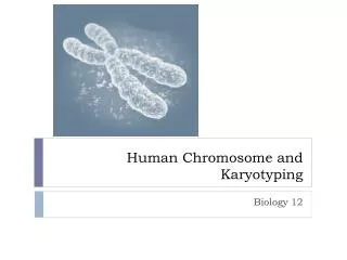
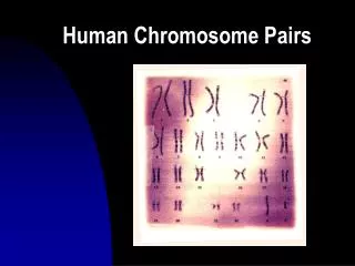
Karyotype Analysis • Preparation of metaphase chromosomes. • To view human chromosomes, geneticists remove white blood cells, stain and photograph their nuclei, then cut the chromosomes from photo with scissors and arrange them in pairs in decreasing size as shown in the next slide.
Changes in the Chromosome Number • Down Syndrome: • People normally have 46 chromosomes: 22 pairs of autosomes and 1 pair of sex chromosomes – XX or XY • In case of Down Syndrome the cell has 47 chromosomes, 3 copies of chromosome 21 – Trisomy 21 • Syndrome is a set of symptoms that occur together.
This condition is characterized by lower than average IQ. Heart malformation, eyelid folds, and others.
Down Syndrome:Trisomy 21 wasfirst described in 1866 by J. Langdon Down
Correlation between maternal age and the incidence of Down syndrome • As women age, the chances they will bear a child with Down syndrome increase
Nondisjunction • Errors in the distribution of chromosomes during meiosis causes changes in the chromosome number. • When homologous chromosomes fail to separate, the 2 chromosomes move toward one pole of the cell, none to the other cell.
Nondisjunction may also affect the sex chromosomes • nondisjunction of the X chromosome creates three possible viable conditions • XXX female • usually taller than average but other symptoms vary • XXY male (Klinefelter syndrome) • sterile male with many female characteristics and diminished mental capacity • XO female (Turner syndrome) • sterile female with webbed neck and diminished stature
Turner Syndrome • A person with one X chromosome and no Y chromosome (XO) is a sterile female. • 2N=45 • This condition is characterized by by folds of skin along the neck, a low hairline at the nape of the neck, and failure to develop adult sexual characteristics at puberty.
Klinefelter Syndrome • A male with 2 X chromosomes and one Y chromosome (XXY) • 2N=47 • Affected people develop as sterile males with small testes, long legs and arms. Most manage well in society
Changes in the Chromosome Structure • Deletion • Inversion • Duplication • Translocation • View “Changes in Chromosome Structure” – animation in my Website
Deletion • The loss of some segments of a chromosome. • Example: a normal sequence of genes on a chromosome is: A B C D E F G H • If this chromosome loses a piece containing genes D E F, it becomes shorter with following sequence: A B C G H
Most deletions are lethal or cause serious disorders. For example, one deletion from human chromosome 5 results in mental retardation. When affected infants cry, they produce sounds rather like a cat’s meow. Hence cri-du-chat (cat-cry), the name of this disorder.
Inversion • A linear stretch of DNA within the chromosome becomes oriented in reverse direction. • Normal sequence of genes is: • A B C D E F G H before damage • A B E D C F G H afterward
Duplication • Gene sequences are repeated many times • Normal sequence of genes before damage • A B C D E F G H • After modification it becomes: • A B C D C D C D C D E F G H or • A B C D E F F F F G H
Translocation • It involves nonhomologous chromosomes • A broken part of a chromosome becomes attached to a nonhomologous chromosome • Sequence of genes in one chromosome is A B C D, and E F G H in the second. • After translocation the sequences will become: A B E F and C D G H
Questions and Answers Q. A chromosome’s gene sequence that was ABCDEFGH before modification and ABCDLMNOPafterward is an example of: A. Translocation Q. A chromosome’s gene sequence that was ABCDEFGH before modification and ABCDCDCDEFGH afterward is an example of: A. Duplication
Questions and Answers Q. A chromosome’s gene sequence that was ABCDEFGH before damage and ABCFG H after is an example of: A. Deletion Q. A chromosome’s gene sequence that was ABCDEFGH before damage and ABFEDCGH after is an example of: A. Inversion