Download
1 / 37
420 likes | 905 Views
Neuromuscular Function. Draw and label a diagram of a motor unit. Terms to know : dendrite cell body (soma) nucleus axon motor end plate synapse. Neuromuscular Function. Draw and label a diagram of a motor unit. Neuromuscular Function.

E N D
Neuromuscular Function Draw and label a diagram of a motor unit. Terms to know: dendrite cell body (soma) nucleus axon motor end plate synapse
Neuromuscular Function Draw and label a diagram of a motor unit.
Neuromuscular Function Draw and label a diagram of a motor unit.
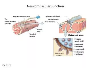
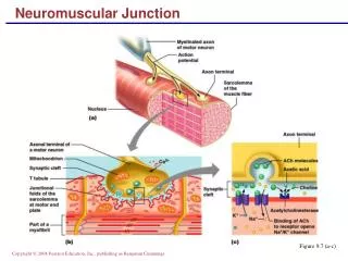
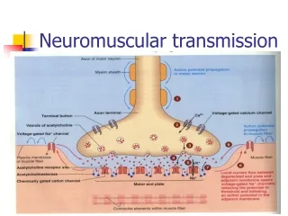
Neuromuscular Function Role of neurotransmitters in stimulating muscle contraction. • Neurotransmitters are chemicals that are used for communication between a neuron at the synapse and another cell.
Neuromuscular Function Role of neurotransmitters in stimulating muscle contraction.
Neuromuscular Function Role of neurotransmitters in stimulating muscle contraction. Acetylcholine (Ach): increases the post-synaptic membrane’s permeability to sodium and potassium ions spreading the impulse over the entire muscle fiber.
Neuromuscular Function Role of neurotransmitters in stimulating muscle contraction.
Neuromuscular Function Role of neurotransmitters in stimulating muscle contraction. Cholinesteraseis an enzyme that catalyzes the hydrolysis of the neurotransmitter acetylcholine into choline and acetic acid, a reaction necessary to allow a neuron to return to its resting state after activation.
Neuromuscular Function Sliding Filament Theory Terms to know: myofibril myofilament sarcomere actin/myosin H zone A band Z line tropomyosin troponin sarcoplasmic reticulum sarcoplasm sarcolemma calcium ions ATP
Neuromuscular Function Sliding Filament Theory Tropomyosin is an actin-binding protein that regulates actin mechanics. Troponin (a protein)is attached to the protein tropomyosin and lies within the groove between actin filaments in muscle tissue. In a relaxed muscle, tropomyosin blocks the attachment site for the myosin crossbridge, thus preventing contraction.
Neuromuscular Function Sliding Filament Theory • The sarcoplasmic reticulum is a special type of smooth ER found in smooth and striated muscle. The only structural difference between this organelle and the smooth endoplasmic reticulum is the medley of protein they have, both bound to their membranes and drifting within the confines of their lumens. This fundamental difference is indicative of their functions: the smooth ER synthesizes molecules and the sarcoplasmic reticulum stores and pumps calcium ions. The sarcoplasmic reticulum contains large stores of calcium, which it sequesters and then releases when the cell is depolarized. This has the effect of triggering muscle contraction.
Neuromuscular Function Sliding Filament Theory • Explains how muscle fibers shorten during a contraction. • When the myosin cross-bridges are activated, they bind with actin, resulting in a conformational change in the cross-bridge, which causes the myosin to tilt and to drag the thin filament toward the center of the sarcomere.
Neuromuscular Function Sliding Filament Theory Steps of a muscle contraction: *Ca++ are released by the sarcoplasmic reticulum. *Ca++ binds to troponin preventing the blocking action of tropomyosin.
Neuromuscular Function Sliding Filament Theory *myosin heads can now attach to active sites on the actin filament. *using ATP, the myosin heads pulls on the actin filament. *myosin head releases the actin when a new ATP is formed.
Neuromuscular Function Sliding Filament Theory
Neuromuscular Function Sliding Filament Theory
Neuromuscular Function Sliding Filament Theory
Neuromuscular Function Sliding Filament Theory
Neuromuscular Function Sliding Filament Theory • Immediately after the myosin head tilts, it breaks away from the active site, rotates back to its original position, and attaches to a new active site farther along the actin filament. Repeated attachments and power strokes cause the filaments to slide past one another, giving rise to the term sliding filament theory. This process continues until the ends of the myosin filaments reaches the Z-disks, or until the Calcium is pumped back into the sarcoplasmic reticulum.
Neuromuscular Function Sliding Filament Theory • During this sliding (contraction), the thin filaments move toward the centre of the sarcomere and protrude into the H-zone, ultimately overlapping. When this occurs, the H zone is no longer visible.
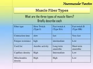
Neuromuscular Function Slow & Fast Twitch Fibers • Slow Twitch: (type 1) *smaller in diameter *reddish color *use aerobic resp. for ATP supply *contain more mitochondria *fire slowly, but take long to fatigue.
Neuromuscular Function Slow & Fast Twitch Fibers Fast Twitch: used for short explosive movements, stop and go sports. Type IIA: *large diameter *white in color *less mitochondria *uses both anaerobic and aerobic energy transfer Type IIB: *same physical characteristics as Type IIA, but strictly uses the glycolytic anaerobic system.
Neuromuscular Function Slow & Fast Twitch Fibers • Slow-twitch, or type I, fibers (sometimes referred to as "Red") have more mitochondria, store oxygen in myoglobin, rely on aerobic metabolism, have a greater capillary to volume ratio and are associated with endurance; these produce ATP more slowly. Marathon runners tend to have more type I fibers, generally through a combination of genetics and training.
Neuromuscular Function Slow & Fast Twitch Fibers • Fast-twitch, or type II, fibers (sometimes referred to as "White") have fewer mitochondria, are capable of more powerful (but shorter) contractions, metabolize ATP more quickly, have a lower capillary to volume ratio, and are more likely to accumulate lactic acid. Weightlifters and sprinters tend to have more type II fibers. Type II fibers are distinguished by their primary sub-types, IIa, IIx, and IIb, as described below.
Neuromuscular Function Slow & Fast Twitch Fibers • Type II fibers come in three primary sub-types, called type IIa, IIx, and IIb. Recent studies show that human skeletal muscle contains type I, IIa, and IIx fibers, though confusingly, human IIx fibers used to be referred to as type IIb. Types IIa, IIx, and IIb fibers are found in skeletal muscle of other mammals (e.g., rodents and cats).
Neuromuscular Function Synovial Joint Movements • Types of Joint Movement: • Abduction: movement away from the body’s center. • Adduction: movement towards the body’s center.
Neuromuscular Function Synovial Joint Movements Circumduction: making circular movements. Dorsiflexion: movement of the ankle elevating the sole. (digging in the heel) Plantar flexion: extending the ankle and elevating the heel. (standing on tiptoes)
Neuromuscular Function Synovial Joint Movements Elevation: occurs when a structure moves in a superior (towards head) manner. Ex. Closing your mouth/elevating the shoulders. Depression: movement is inferior (towards feet). Ex. opening your mouth/lowering the shoulders
Neuromuscular Function Synovial Joint Movements Extension: movement that increases the angle between articulating elements opening the joint. Flexion: decreases the angle between articulating elements and closes the joint.
Neuromuscular Function Synovial Joint Movements Pronation: rotating the palm down. Supination: rotating the palm up. Rotation: turning the body around a longitudinal axis.
Neuromuscular Function Synovial Joint Movements Inversion: when the ankle rolls outward. Eversion: ankle roles inward.
Neuromuscular Function Types of muscle contraction • Isotonic: Increase in loadson muscles. (concentric and eccentric muscle actions.) • Concentric: muscle is shortened during contraction. • Eccentric: muscle is contracting while lengthening.
Neuromuscular Function Types of muscle contraction Isometric: muscle generates force without changing length. Ex. Hand grip, plank position & carrying a bag. Isokinetic: the speed of movement is fixed and the resistance varies with the force exerted. *requires special equipment!
Neuromuscular Function Reciprocal Inhibition Describes muscles on one side of a joint relaxing while the other side is contracting. (antagonistic pairs) • Agonist: muscle that causes the movement. • Antagonist: muscle that works opposite the agonist to return the joint to its initial position.
Neuromuscular Function Movement Analysis Describe movement considering the following terms: Abduction Adduction Circumduction Dorsiflexion Plantar flexion Elevation Depression Extension Flexion Pronation Supination Rotation Inversion Eversion Isotonic Concentric Eccentric Isometric Isokinetic muscle & exercise directory
Neuromuscular Function Delayed onset muscle soreness (DOMS) The pain and stiffness felt in muscles several hours to days after unaccustomed or strenuous exercise. *brought on by eccentric contractions of the muscle causing pressure at the nerve endings. Read the article and summarize http://sportsmedicine.about.com/cs/injuries/a/doms.htm