Download
1 / 17
200 likes | 394 Views
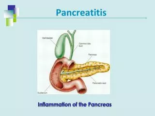
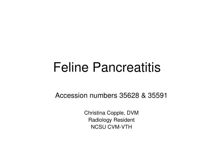
Feline Pancreatitis. Accession numbers 35628 & 35591 Christina Copple, DVM Radiology Resident NCSU CVM-VTH. Small, thin, elongate, V-shaped Two lobes and a body Left lobe Body Right lobe. General Anatomy. *Left of midline and parallel to spine. General Anatomy.

E N D
Feline Pancreatitis Accession numbers 35628 & 35591 Christina Copple, DVM Radiology Resident NCSU CVM-VTH
Small, thin, elongate, V-shaped Two lobes and a body Left lobe Body Right lobe General Anatomy *Left of midline and parallel to spine
Left lobe between greater curvature of stomach and transverse colon extending towards cranial pole of left kidney along medial aspect of spleen Body within the cranial duodenal flexure, between pylorus and proximal descending duodenum Right lobe extends caudally along the medial (mesenteric) aspect of the descending duodenum and lateral to the ascending colon Normal pancreatic thickness should measure <15 mm Ultrasound Anatomy *This is a canine diagram, but orientation of the pancreas remains the same with the stomach more leftward and parallel to spine
Pancreatitis = inflammation of the pancreas due to autodigestion Feline pancreatitis often more chronic with severe or mild forms Triaditis = cholangiohepatitis & pancreatitis & inflammatory bowel disease Feline Pancreatitis
Several Types that require histopathology to definitively differentiate Acute Necrotizing Acute Suppurative Chronic Nonsuppurative Causes Idiopathic (most common) Infections (T. gondii, A. pseudofelineus, panleukopenia and FIP) Blunt Trauma Aggressive surgical manipulation Ischemia Inflammatory diseases OP toxicity Drug? Feline Pancreatitis
Radiography increased, irregular soft tissue opacity with focal loss of serosal detail in right mid-cranial abdomen rightward or ventral displacement of proximal descending duodenum = creating a broad curve also called the “C” sign Descending duodenum may be gas-distended May have caudal displacement of transverse colon Depending on chronicity - mineralized foci in the pancreatic region MAY BE NORMAL Ultrasonography Enlargement of pancreas with irregular and ill-defined margins, appearing hypoechoic or complex Surrounding mesentery is hyperechoic and there may be local peritoneal fluid = indicating a focal peritonitis Pain expressed when applying pressure to image the region of the pancreas MAY BE NORMAL Diagnostic Imaging *Normal findings do not rule-out pancreatitis!
Sugar 9yr old FS DSH 3 day hx of vomiting, lethargy, anorexia Possible foreign body ingestion 3 weeks ago CBC: marked inflammatory leukogram with left shift CHEM: elevated ALP/ALT/Tbili, stress hyperglycemia Felv and FIV negative Accession numbers 35628 & 35591 http://photo.xanga.com/Bethberry/dc657188229127/photo.html
Accession number 35591 Left Lobe Peritoneal Effusion Right Lobe
LITERATURE REVIEW Head, et al. Evaluation Of The Feline Pancreas Using Computed Tomography And Radiolabeled Leukocytes. VetRadUS 2003. • CT – single most important imaging modality in humans to evaluate the pancreas; to diagnose pancreatitis in humans: sensitivity = 92%, specificity = 100% • Radiolabeled Granulocyte Scintigraphy – detect abdominal inflammation in humans and horses; Hexamethylpropyleneamine oxime (HMPAO) attached to 99mTC effectively labels granulocytes and localizes in areas of neutrophil accumulation = acute inflammation • Six, healthy, 8mth old M cats, normal CBC/Chem/amylase/lipase/TLI/abdominal rads/abdominal ultrasound • Histopathology of pancreatic tissue in all cats confirmed absence of pancreatic inflammation
Nuclear Scintigraphy: labeled radiopharmaceutical uptake predominantly in lung, spleen, and liver in order of decreasing activity, some intestinal and renal/urinary activity NO APPRECIABLE UPTAKE IN REGION OF PANCREAS LITERATURE REVIEW
CT: pancreas easily identified, pre-contrast hypoattenuating (mean HU-37.45) to liver and spleen with scalloped sharply defined margins and the peripancreatitc fat was hypoattenuating to the pancreas itself, peak contrast enhancement immediately was uniform but still less attenuating when compared to post-contrast spleen, wash out was gradual over 30 minutes LITERATURE REVIEW
LITERATURE REVIEW Head, et al. Use Of Computed Tomography And Radiolabeled Leukocytes In A Cat With Pancreatitis. VetRadUS. 2005. • Case study: 9yr old MC DLH, anorexia, depression and occasional vomiting X 5 days, results of laboratory tests were non-specific • Abdominal rads- loss of serosal detail in central/cranial abdomen • Abdominal US- enlarged, hypoechoic, less homogenous pancreas, trace peri-pancreatic fluid, surrounding mesentery was hyperechoic, regional perilytic ileus in descending duodenum • No response to supportive care and appropriate medical care over 13 days, decided for exploratory laparotomy with biopsies and jejunostomy tube placement, owner consented to granulocyte labeling procedure and CT prior to SX • Histopathology diagnosed moderate-to-severe pancreatitis
Nuclear Scintigraphy: 99mTC-HMPAO labeled leukocytes were injected IV, utilized previously reported protocol but additional images were acquired at 17hrs post-injection with 420,000 counts instead of 500,000 Same previous distribution in lung, liver, and spleen but abnormal uptake in region of right pancreatic lobe and body LITERATURE REVIEW
CT: utilized previously published protocol used except this patient was administered Iohexol instead of Conray, larger and irregularly marginated with heterogeneous attenuation and multiple small hypoattenuating foci, linear hyperattenuating streaks in the surrounding mesentery, peak contrast enhancement was not immediate and took 10 minutes to occur with a normal washout period LITERATURE REVIEW
References • Thrall, Donald E., DVM, PhD, DACVR. Textbook of Veterinary Diagnostic Radiology. 5th ed. Saunders. St. Louis, MO, 2007. • Nyland, Thomas G., DVM, DACVR and John S. Mattoon, DVM, DACVR. Small Animal Diagnostic Ultrasound. 2nd ed. Saunders. Philadelphia, PA, 2002. • August, John R., BVetMed, MS, MRCVS, DACVIM editor. Consultations in Feline Medicine. Volume 5. Elsevier. St. Louis, MO, 2006. • Ettinger, Stephen J., DVM and Edward C. Feldman, DVM. Textbook of Veterinary Internal Medicine Diseases of the Dog and Cat. 6th ed. Volume 2. Elsevier. St. Louis, MO, 2005. • Evans, Howard E., Ph.D. and Alexander deLahunta, D.V.M., Ph.D. Guide To The Dissection Of The Dog. 5th ed. W.B. Saunders. Philadelphia, PA, 2000. • Dyce et al. Textbook Of Veterinary Anatomy. 2nd ed. W.B. Saunders. Philadelphia, PA, 1996. • Head, Laurie L., et al. Evaluation Of The Feline Pancreas Using Computed Tomography and Radiolabled Leukocytes. VetRadUS. Vol. 44. No. 4, 2003, pp 420-428. • Head, Laurie L., et al. Use Of Computed Tomography And Radiolabeled Leukocytes In A Cat With Pancreatitis. VetRadUS. Vol. 46. No. 3, 2005, pp 263-266.