Download
1 / 25
260 likes | 589 Views
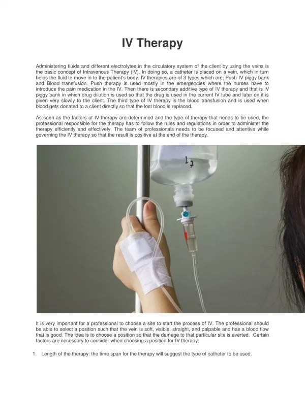
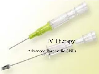
IV therapy PART 2. Catherine Luksic BSN, RN. Methods of iv infusion. Primary infusion “maintenance infusion” “continuous infusion” Via gravity Via electronic pump Secondary infusion “piggyback” “IVPB” Usually via electronic pump. Equipment: IV tubing. Primary IV administration set

E N D
IV therapyPART 2 Catherine Luksic BSN, RN
Methods of iv infusion • Primary infusion • “maintenance infusion” • “continuous infusion” • Via gravity • Via electronic pump • Secondary infusion • “piggyback” • “IVPB” • Usually via electronic pump
Equipment: IV tubing • Primary IV administration set • Gravity infusion • Electronic pump infusion • Secondary IV administration set • “piggyback tubing” • Blood administration Y set • Extension tubing • Intermittent infusion lock • HL or SL
Methods of IV Infusion • Gravity Drip • Manually set, regulated w/ roller clamp • Simplest method • Count drops manually • Macrodrip tubing - drop factor determined by manufacturer • Can range from 10-20 gtts/mL(common 10gtts/mL) • standard primary tubing • for rapid infusions • Microdrip tubing - 60gtts/mL • Used for peds, elderly, slower rates
Gravity Infusion • Advantages: • Easy • Does not require power source or pump • Can set-up quickly • Disadvantages: • Not greatly accurate • No free-flow prevention • Room for error
Calculating a gravity IV rate • Volume ordered to be infused • 1000 ml or 1000 cc • Drop factor of tubing being used • Rate of infusion as per order • 100 ml /hr (or 100 cc/hr) **refer to IV calculations worksheets
Rate of IV INFUSION • Check physician orders ! • Gravity drip or electronic pump ? • cc/hr = ml/hr • KVO (10-20 ml/hr) • TKO • Check IV site & IV rate at least every hour
Priming • Refer to procedure • Maintain sterility • Remember to close the roller clamp 1st ** • ?? Invert all Y-sites and tap to remove air • Demonstration • Practice
Factors that will affect flow rate • Height of bag • 36 inches above heart • Position of roller clamp • is it open or closed ? • Patency of tubing • Check for kinks
Slowed Infusion Rate • Check rate - has it been changed? • Check tubing - is it kinked? • Check clamp(s) - are they open? • Check site - • is cath or vein being compressed? • ANY abnormality? • Look for sign of infiltration
Electronic Control Devices • Purpose – improve accuracy of delivery. • Requires power source • Deliver a preset fluid rate over a specified period • Uses constant force • Always use pump w/ TPN, central lines, titrated medications, blood products
Electronic Pump Alarms(Common) • Occlusion • Air-in line • Infusion complete • Power
Electronic IV Pumps • SINGLE CHANNEL • MULTI-CHANNEL • PCA (Patient controlled analgesia) • AMBULATORY IV PUMPS • Home care use
NURSING PROCESS • ASSESSMENT • DIAGNOSIS • PLANNING • IMPLEMENTATION • EVALUATION
ASSESSMENT • FIRST STEP, AS ALWAYS • chronic conditions • use of long-term medications (anticoagulants) • previous IV experiences/problems • allergies (especially latex & antimicrobial agents) • hand dominance • Skin *Cultural considerations, communication barriers, level of understanding
Assessment • Physical exam • Neurological status – AAO, ability to understand and cooperate • Cardiovascular status – color, pulses, edema, appearance of veins • Skin – bruising, rashes, lesions
NURSING DIAGNOSIS • Examples: • Risk for injury related to (lack of knowledge regarding equipment) • Knowledge deficit related to (new IV insertion) AEB (pt verbalization …) • Impaired physical mobility related to placement of peripheral IV AEB… • Anxiety related to (initiation of IV therapy) AEB... • Alteration in comfort: Pain
Planning • Patient outcomes and goals - what do you (and the patient!) expect. • Ex: Pt. will remain free of S/S of complications related to IV therapy • More specific – Pt. will remain free of signs of phlebitis
Implementation • Nursing Care: • Check site HOURLY for complications - redness, pain, edema, infiltration • Instruct pt. to call nurse immediately: pain, bleeding, other concerns. • Instruct pt. to call nurse if pump alarm sounds. • Maintain correct infusion rate as ordered. • Secure IV (to prevent accidental dislodging of catheter during movement). • Change tubing according to hospital policy (usually q 72 hr.) • Change IV site according to hospital policy (usually q 72 hrs.)
Implementation • Nursing Care: • 2011 Infusion Nurse Society (recommendations) • Do not change IV tubing more frequently than q 96 hrs. • If IVPB tubing is detached from continuous tubing, change q 24 hrs.
Evaluation • How will you know if the goal/outcome has been achieved? • Assessment • Patient record • Lab values • Communication