Download
1 / 17
180 likes | 443 Views
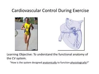
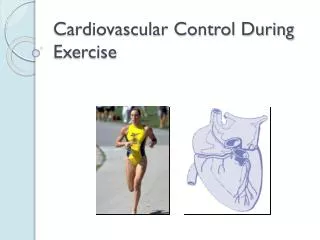
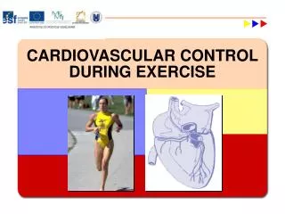
Cardiovascular Control During Exercise. Cardiovascular Functions. Delivery Oxygen and nutrients Removal CO2 and metabolic wastes Transport hormones Maintenance Body temperature Fluid leves and pH Prevention infection. The Heart. Blood flow through the heart (fig 8.1) The myocardium

E N D
Cardiovascular Functions • Delivery • Oxygen and nutrients • Removal • CO2 and metabolic wastes • Transport • hormones • Maintenance • Body temperature • Fluid leves and pH • Prevention • infection
The Heart • Blood flow through the heart(fig 8.1) • The myocardium • interconnected cardiac muscle • hypertrophy of left ventrical • The cardiac conduction system(fig 8.3) • Autoconduction: the ability to generate its own electrical signal rythmically without neural stimuation. • SA node: (pacemaker) sends the electrical impulse to the atria and reaches the AV node. • AV node: conducts the impulse from the atria into the ventricals through the .... • AV bundle and Perkinji fibers where it travels along the septum and to the ventrical walls starting at the Apex.
The Heart • Extrinsic control of heart activity • the parasympathetic nervous system • decreases H.R. & force of heart contraction • the sympathetic nervous system • increases H.R. & force of heart contraction • the endocrine system: release norepinephrine and epinephrine to increase H.R. • The ECG(fig 8.4) • records the electrical activity of the heart • the P wave: atrial depolarization • the QRS complex: ventricular depolarization • the T wave: ventricular repolarization
Cardiac Arrhythmias • Bradycardia: “slow heart” • Resting H.R. < 60 • Tachycardia: “fast heart” • Resting H.R. > 100 • Symptoms include • Fatigue • Dizziness • Lightheadedness • Fainting • Premature ventricular contraction: “skipped beat” • Ventricular Fibrillation: “uncoordinated beat”
The Heart • The Cardiac Cycle: includes all of the events between two consecutive cycles • Diastole: relaxation phase • Systole: contraction phase • Stroke Volume(SV):the amount of blood ejected from the left ventrical (fig 8.5). • SV = EDV - ESV • end diastolic volume (EDV) • end systolic volume (ESV) • ejection fraction (EF) = (SV / EDV) X 100% • cardiac output (Q) = HR X SV
The Vascular System • Method: Aorta --> Arteries --> Arterioles --> Capillaries -->Venuoles --> Veins --> Vena Cava • Coronary arteries • Return of blood to the heart • breathing increases thoracic pressure • muscles create a pumping action • valves prevent backflow
The Vascular System • Distribution of blood(fig 8.6) • autoregulation: the vessels ability to detect the local chemical changes and regulate its own blood flow to meet the needs of the tissues. • extrensic neural control: regulated largely by the sympathetic nervous system by constricting blood vessels of lesser need. • redistribution of venous blood: creating more available blood to meet the needs of the body. • During Exercise: blood is redirected to the areas where it is needed most • Muscles receive up to 80%
The Vascular System • Redistribution of Venous Blood • 64% of blood pools in the veins waiting for the need. • Blood pressure • systolic / diastolic • Measured sitting and supine/prone • control: weight loss, diet, exercise, med’s • Hypertension: 140 / 100 • Hypotension: 100 / 60
The Blood • Functions • Transportation of nutrients, hormones, etc. • Temperature regulation • Maintain (pH) balance • Blood volume and composition • Men 5 - 6 L, Women 4 - 5 L • composition (fig 8.8) • 55% plasma • 90% water • 45% hematocrit • red blood cells: transport oxygen primarily bound to their hemoglobin (iron). • White blood cells • platelets
The Blood • Blood viscosity:refers to the thickness of the blood. • increased viscosity restricts blood flow but increases oxygen carrying capacity. • decreased viscosity increases blood flow but decreases oxygen carrying capacity.
Cardiovascular Response to Exercise • Increased stroke volume(fig 8.11) • only up to 40%-60% of maximal capacity & then plateaus (caused by reduced filling time at higher h.r. ?) • increased volume of venous blood return • increased muscle pumping of venous blood • increased breathing (thoracic pressure) • supine positions • increased ventrical enlargement capacity • Frank-Starling law: when the ventricle stretches more, it will contract with more force. • increased ventrical contractility • aortic or pulmonary artery pressure
Cardiovascular Response to Exercise • Increased heart rate / cardiac output (fig 8.10) • Anticipatory response(increased heart rate before exercise) • Caused by the release of epinephrine • Steady state heart rate: during steady exercise • Maximum heart rate= 220 - age
Cardiovascular Response to Exercise • Redistribution of blood to the working muscles by reducing blood flow to the kidneys, stomach, liver and intestines. • Redistribution of blood to the skin in order to maintain body temperature. • Increased metabolic rate of working muscles • Autoregulation is triggered by low muscle Po2 • Cardiovascular drift: increased H.R. compensates for a decreased S.V. from a decreased total blood volume to maintain Q. • redistribution • decreased blood plasma
Cardiovascular Response to Exercise • Systolic B.P. increases with intensity • valsalva during resistance exercise • increased use of upper body musculature • Diastolic B. P. does not change
Cardiovascular Responses to Exercise • Increased A-V O2 difference: representing the amount of O2 extracted from the blood to be used by the muscles. • Decreased plasma volume =decreased performance increased blood pressure forces water from the vascular system to the interstitial spaces. • increased intramuscular osmotic pressure attracts fluid to the muscles. • sweating • Increased blood viscosity • decreasing O2 transport • Decreased blood pH level