Download
1 / 81
871 likes | 1.32k Views
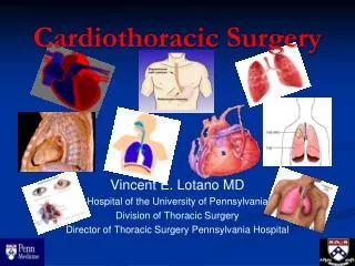
COMPLICATIONS OF CARDIOTHORACIC SURGERY. Jacqueline Palmer-Powell, RN Nurse Educator/CNS. Complications Commonly Resulting From Cardio-Thoracic Surgery. CVA. CVA. Devastating complication that results in lasting deficits of death.

E N D
COMPLICATIONS OFCARDIOTHORACICSURGERY Jacqueline Palmer-Powell, RN Nurse Educator/CNS
Complications Commonly Resulting From Cardio-Thoracic Surgery
CVA • Devastating complication that results in lasting deficits of death. • Other neurological complications which are more subtle occur with more frequency. • Incidence • -CABG with CPB: 2-5% • -Valve Surgery: 5-10%
Risk Factors • Prior history of stroke, HTN, DM • Carotid disease, carotid bruit • Advanced age • Atrial Fib
Diagnosis & Treatment • CT Scan demonstrates cerebral infarct within 1-2 days • No treatment exist other than palliative • Early rehabilitation • Family counseling
Hypotension • Definition: Systolic BP<100 • Cause: hypovolemia, excessive vasodilation, sepsis, elevated or decreased HR • Treatment: identify cause!, volume replacement, use of IV drips
Contributing Factors in Bleeding Complication • Pre-op Post-op • Acute MI treated with - Vigorous chest thrombolytics (failed) tube stripping • Aspirin - Hypertension • IIBIIIA Platelet - Heparin • Co-morbid states (uremia, liver disease)
What Constitutes Excessive Bleeding? • Chest Tube Drainage: • - >500cc/hr in first hour • - >400cc/hr during first 2 hours • - >300cc/hr during first 3 hours • - >200cc/hr during first 6 hours
Basis of Coagulation • Coagulation Cascade with the help of endothelium & platelets is the body’s defense to minimize blood loss. • A vascular insults stimulates formation of platelet plug thru platelet activation, adhesion & aggregation. The plug is then stabilized thru clotting cascade to a fibrin clot
Effect of CPB on Coagulation • Major CPB induced coagulopathy results from platelet activation, dysfunction & destruction. • Structural damage to platelets & RBC’s can occur thru shearing forces & turbulence in CPB pump, circuits & suction devices
Preventing Bleeding Before Surgery • Complete History • - Questions about previous surgery, family history, bruising, heavy menses • - medication history (Prescribed, OTC & herbal) • Physical Exam • Blood Work
Pre-op Bleeding Prevention • Identification of patient with co-morbid states that may contribute to bleeding: • - Uremia: Causes platelet dysfunction thru impaired VWF interaction with platelets. • - Acute liver dysfunction: may result in factor deficiency as a results of impaired factor production & may lead to DIC
Drugs Which Affect Bleeding • Aspirin • NSAIDS • IIBIIIA Platelet Inhibitors • Coumadin • Thrombolytics • Heparin
Prevention & Treatment of Bleeding in OR • Thorough search for bleeding before chest closure including careful inspection of skin, sternum, suture sites • Autotransfusion: pre-op blood donation (self-directed) with re-infusion after CPB • Cell-saver-blood drained from chest tubes in OR collected thru special filters & reinfused after surgery
DIC • Diagnosis: Increased products of fibrin degradation (d-dimer), thrombocytopenia & prolongation of both PT & PTT • Treatment: Replacement with PRBC, FFP & platelets. If fibrinogen level low, replacement with cryoprecipitate is preferable. Drugs (Amikar, Aprotinin) may be useful in treating DIC. High mortality!
Drugs Used To Treat Bleeding • Protamine SO4: Protein derived from salmon sperm. Used to neutralize effects of heparin • DDAVP: Analog of vasopressin may be used when a patient continues to bleed despite normal coagulation profile & platelet counts • Antifubrinolytics: Help to achieve homeostasis in patients with excessive fibrinolysis
Blood Products • Depending on patient presentation, history & lab results the bleeding may require infusion(s) of PRBC, platelets, FFP, &/or cryoprecipitate to control the bleeding & prevent hemodynamic instability
Re-Op for Bleeding Chest Exploration • <3% of patients require re-op to search for bleeding • Bleeding causing tamponade or severe hypotension requires immediate re-op • Coagulopathy must be distinguished from anatomic cause
Signs & Symptoms of LCOS • Results directly from inadequate tissue perfusion & increased sympathetic activity. • Cool, clammy skin with slow capillary refill • Oliguria • Mental status changes • Metabolic acidosis • Fall in SVO2
Causes of LCOS • Any pre-op condition - Post –op conditions that that causes impairment cause myocardial dysfunction: of preload, after load &/or contractility - hypothermia • Events in OR - acidosis • Arrhythmias - hypercarbia • Inadequate preload or - volume overload elevated intrathoracic - increased afterload pressure
Treatment of LCOS • Heart rate manipulation • Preload • Afterload • Myocardial Contractility
Shock • Clinical syndrome representing an extreme state of circulatory failure • Impaired tissue perfusion leading to cellular dysfunction • Complex group of signs & symptoms that can be caused by a variety of factors
Clinical Manifestations of Shock • Directly related to pathophysiologic mechnaisms are involved. Progression is variable & depends on: • -Patient age & prior state of health • Duration of shock state • Response to treatment • Correction of treatable cause
3 Stages of Shock • Early or compensatory • Intermediate or progressive • Late or irreversible
Classification • Vascular Tone (Distributive) • Neurogenic • Septic • Anaphylactic • Intravascular Volume • Hypovolemic • Ability of heart to act as pump • Cardiogenic
Hemodynamic Changes CO CVP SVR PAP PAWP Distributive ↑or↑ ↓or↑ ↓or↑ ↓or↑ ↓or↑ Hypovolemic ↓ ↓ ↑ ↓ ↓ Cardiogenic ↓ ↑ ↑ ↑ ↑
Treating Shock States • Position: Supine/let elevation (if possible) • Trendelenberg should be avoided: • Initiates aortic & carotid sinus reflex • Impaired cerebral blood flow • Decreased filling of coronary arteries
Fluids • Shock almost always involves a decrease in effective circulating volume • Need for volume expansion • Fluid challenge
Septic Shock • Occurs in patients as a result of overwhelming infection • More common in infants, elderly & immuno-compromised • Clinical presentation can be subtle in elderly, debilitated or malnourished patients
Warm Shock • Vasodilation → ↓SVR ↑ or normal CO • BP ↓ but skin is pink, warm & dry • Urine output is adequate
Cold Shock • Vasoconstriction ↓ ↑ SVR • ↓ CO • ↓ BP • ↓ Urine output • Metabolic Acidosis
Hypovolemic Shock • Loss of intravascular volume ↓ • Decreased venous return to heart ↓ • Circulatory insufficiency ↓ • Inadequate tissue perfusion
Cardiogenic Shock • Pump failure • Occurs when the heart can no longer efficiently pump blood. CO is significantly decreased • Major cause: extensive myocardial injury secondary to MI
Treatment of Cardiogenic Shock • Treat reversible cause • Goal of treatment is to: • Increase Cardiac Contractility • Decrease Afterload (workload) • Careful fluid replacement (if needed) • IABP insertion • Drug therapy
Cardiac Performance Low CO Cardiogenic Shock Dobutamine Vasodilator LABP Dopamine Preload ↓ ↓ ↓ ↑ Contractility↑↑ - - ↑ Afterload ↓ ↓↓ ↓ ↑ Heart Rate - - - ↑ MVO2 - ↓↓↓ ↓↓ ↑↑↑↑
Treatment of Peri-Op/Post-OpIschemia • Evaluate/Investigation of cause • Drugs: • Belta Blockers • Nitrates • Vasopressors • Calcium Blockers
Atrial Fibrillation • Nearly 30% of patients undergoing coronary surgery & up to 50% of patients with valvular disease develop AF • Occurs in up to 5% of patients afterr any major surgery
Myocardial Ischemia • Ischemia results from an imbalance between myocardial O2supply & demand. • Can be due to: • ↑ Demand • ↓ Supply • Coronary Vasospasm
Causes of Ischemia Post-Op • Incomplete mycardial protection during aortic cross-clamp, incomplete revascularization, vasospasm, atheromatous emboli, thrombosis of native vessel or new graft • Myocardial revascularization patients are at higher risk of peri-op infarct than other CT surgery patients
Causes of Post-Op A-Fib • Common Causes Other Causes • Electrolyte problems - Valve surgery • Advanced age - History of RF • Hypervolemia - Duration of x-clamp • CHF -CPB • D/C of pre-op meds -Cardioplegia method • Hypoxia -Sepsis • ETOH abuse
Treatment of AF • Prompt identification & treatment of cause • Chemical cardioversion • First give drugs to treat rate • Then drugs to convert to SR • Synchronized cardio version
Brady Arrhythmias • Sinus Bradycardia • Heart Blocks • Cause: • Overuse of Beta blockers pre-op • Manipulation or destruction of SA or AV nodes • Hypoxia • Vagal stimulation
Treatment • Treat Cause • Pacing • Drugs