Download
1 / 18
180 likes | 436 Views
Testicular Ultrasound. Normal and Pathologic Appearances. Indications for Testicular Ultrasound. Assessment of palpable abnormalities Breeding soundness examination Assessment of painful or swollen testes Routine assessment of apparently normal intact males. Normal US Appearance.

E N D
Testicular Ultrasound Normal and Pathologic Appearances
Indications for Testicular Ultrasound • Assessment of palpable abnormalities • Breeding soundness examination • Assessment of painful or swollen testes • Routine assessment of apparently normal intact males
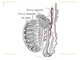
Normal US Appearance • Coarse, homogeneous echotexture • Central, hyperechoic mediastinum testis—2 mm on sagittal and transverse sections • Occasional hyperechoic flecks • Tail of epididymis—hypoechoic to anechoic • Should be bilaterally symmetric—should vary by less than 4.5 mm Pugh CR, Konde LJ, Park RD. Testicular ultrasound in the normal dog. Veterinary Radiology, 31(4), 1990: 195-199.
Normal Examination • Use a high frequency transducer or standoff pad (we use the 13-5 probe) • Sagittal plane • Transverse plane • Dorsal plane—good for imaging epididymis Pugh CR, Konde LJ, Park RD. Testicular ultrasound in the normal dog. Veterinary Radiology, 31(4), 1990: 195-199.
“Winston” • 7 year old male Mastiff • Presented for enlarged prostate • Ultrasound findings included prostatomegaly and a small left testicular nodule
Testicular Diseases • NEOPLASIA • Hernia • Hydrocele—compromised lymphatics (e.g. LSA), hernia, infarction, neoplasia, idiopathic • Torsion—often in retained/tumorous testes—US is a good early diagnostic (before permanent damage) • Hematocele—trauma, neoplasia, DM • Pyocele (infectious)—Blasto, RMSF(?) • Atrophy—contralateral tumor, temp, trauma • Cryptorchidism Sonographic evaluation of canine testicular and scrotal abnormalities: a review of 26 case histories. Vet Rad. 1991: 243-250.
Testicular Neoplasia • Sertoli Cell Tumor • Interstitial Cell Tumor • Seminoma Sonographic evaluation of canine testicular and scrotal abnormalities: a review of 26 case histories. Vet Rad. 1991: 243-250.
Sertoli Cell Tumor • From sustentacular cells of seminiferous tubules • Firm, lobulated, white to grey, often large, greasy (eew!) • May show clinical signs of feminization • More common in cryptorchid testes McEntee MC. Reproductive Oncology. Clinical Techniques in SA Practice. Aug 2002. 138-143.
Sertoli Cell Tumor (cont’d) • Mean age 9.5 yo (younger for cryptorchid dogs) • 9% metastasis—MILN, other LNN, rarely spleen, liver, kidney • Rare in cats (2 reports) • No characteristic US findings (although one must wonder if size could be suggestive) McEntee MC. Reproductive Oncology. Clinical Techniques in SA Practice. Aug 2002. 138-143.
Interstitial Cell Tumor • Leydig cells between seminiferous tubules • Small, discrete, non-palpable (majority under 2 cm) • Soft, bulging, bright yellow or orange (!) • Often cystic • Always scrotal • Metastases very rare McEntee MC. Reproductive Oncology. Clinical Techniques in SA Practice. Aug 2002. 138-143.
Interstitial Cell Tumor (cont’d) • Increased testosterone productionprostatic disease, perianal gland neoplasia, perineal hernia more common • Rare in cats (1 report—incidental finding) • No characteristic US findings McEntee MC. Reproductive Oncology. Clinical Techniques in SA Practice. Aug 2002. 138-143.
Seminoma • Germinal epithelium of seminiferous tubule • Homogeneous, soft, bulging, cream colored • Often large (1 mm to 10 cm diameter; 75% under 2 cm) • Associated with cryptorchidism • Rare metastasis—9%. Usually to MILN, other LNN, occasionally lung • Rare in cats—1 report • No characteristic US appearance—size?? McEntee MC. Reproductive Oncology. Clinical Techniques in SA Practice. Aug 2002. 138-143.
Testicular Tumors—More Fun Facts • ANY can show signs of feminization, Sertoli most common • Majority are asymptomatic—may affect fertility • Many Vietnam-era MWD’s had seminomas (herbicide/pesticide exposure??) • Syndrome in middle-aged, miniature Schnauzer cryptorchid male pseudohermaphrodites—Sertoli cell tumor produces estrogen and causes pyometra or mucometra of the remnant uterus!! (6 dogs) McEntee MC. Reproductive Oncology. Clinical Techniques in SA Practice. Aug 2002. 138-143.
Staging • Abdominal and testicular ultrasound • Thoracic rads (mets rare) • Serum testosterone/estrogen/progesterone • Abdominal rads (look for masses—retained testes) • CBC—look for bone marrow suppression • Coagulation profile (if anemia, petechiae, hemorrhage) McEntee MC. Reproductive Oncology. Clinical Techniques in SA Practice. Aug 2002. 138-143.
Treatment • Castration usually curative • Cisplatin has been used • Radiation for seminoma metastatic to sublumbar lymph nodes McEntee MC. Reproductive Oncology. Clinical Techniques in SA Practice. Aug 2002. 138-143.