Download
1 / 37
370 likes | 447 Views
Chapter 11. How Cells Reproduce. S phase (DNA synthesis; chromosome duplication). Interphase: metabolism and growth (90% of time). G 1. G 2. Mitotic (M) phase: cell division (10% of time). Cytokinesis (division of cytoplasm). Mitosis (division of nucleus). Figure 8.6.

E N D
Chapter 11 How Cells Reproduce
S phase (DNA synthesis; chromosome duplication) Interphase: metabolism and growth (90% of time) G1 G2 Mitotic (M) phase: cell division (10% of time) Cytokinesis (division of cytoplasm) Mitosis (division of nucleus) Figure 8.6
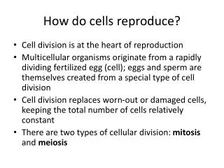
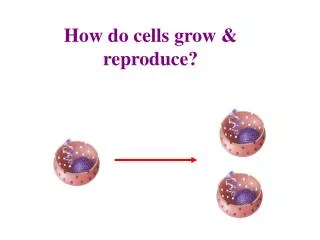
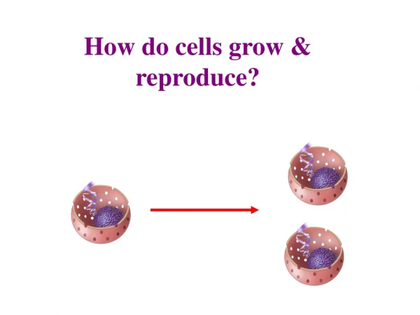
11.1 How Do Cells Reproduce? • Multiplication by division • A life cycle is a collective series of events that an organism passes through during its lifetime • Even single cells of multicellular organisms have a life cycle • Cell cycle: a series of events from the time a cell forms until its cytoplasm divides
Multiplication by Division (cont’d.) • A typical cell spends most of its life in interphase: interval between mitotic divisions when a cell grows • During interphase, a cell roughly doubles the number of its cytoplasmic components, and replicates its DNA
Multiplication by Division • Mitosis: process of nuclear division that maintains chromosome number • Mitosis and cytoplasmic division are the basis of: • Developmental processes (e.g., increases in body size and tissue remodeling) • Replacement of damaged or dead cells • Eukaryotic asexual reproduction (offspring are produced by one parent)
Multiplication by Division • When a cell divides by mitosis, it produces two descendant cells • Each with the same number and type of chromosomes as the parent • Human body cells are diploid (contain pairs of chromosomes) • With exception, the chromosomes of each pair are homologous: have the same length, shape, and genes
Multiplication by Division • During mitosis, the sister chromatids are pulled apart • Each sister chromatids end up in separate nuclei that are packaged into separate cells
Multiplication by Division A Pair of homologous chromosomes in a cell during G1. Both are unduplicated. B By G2, each chromosome has been duplicated. C Mitosis and cyto-plasmic division package one copy of each chromosome into each of two new cells.
Control Over the Cell Cycle • Whether or not a cell divides is determined by mechanisms of gene expression control • “Brakes” on the cell cycle normally keep the vast majority of cells in G1 • “Checkpoint genes” monitor: • The completion of DNA copying • DNA damage • Nutrient availability
11.2 What Is the Sequence of Events During Mitosis? • Before mitosis: • Interphase: chromosomes are loosened to allow transcription and DNA replication • Early prophase: in preparation for nuclear division the chromosomes begin to pack tightly
What Is the Sequence of Events During Mitosis? • Prophose (first stage of mitosis): • Chromosomes further condense • One of the two centrosomes move to the opposite end of cell • Microtubules assemble and lengthen, forming a spindle (functions to move chromosomes) • Nuclear envelope breaks up • Sister chromatids are attached to opposite centrosomes
What Is the Sequence of Events During Mitosis? spindle pole
What Is the Sequence of Events During Mitosis? • Final stages of mitosis: • Metaphase: all chromosomes are aligned midway between spindle poles • Anaphase: sister chromatids separate and move toward opposite spindle poles • Telophase: chromosomes arrive at opposite spindle poles and decondense; two new nuclei form
11.3 How Does a Eukaryotic Cell Divide? • In most eukaryotes, cytokinesis (cytoplasmic division) occurs between late anaphase and the end of telophase • The process of cytokinesis differs between plant and animal cells
How Does a Eukaryotic Cell Divide? • Animal cell cytokinesis: • Typical animal cells pinch themselves in two after nuclear division ends • The spindle begins to disassemble during telophase • Contractile rings drag the plasma membrane inward • Cleavage furrow (indentation) forms
How Does a Eukaryotic Cell Divide? • Plant cell cytokinesis: • Dividing plant cells face a challenge because a cell wall surrounds their plasma membrane • By the end of anaphase short microtubules form on either side of the future plane of division • Disk-shaped structure called the cell plate forms and eventually partitions the cytoplasm • The cell plate forms into two new cell walls
11.4 What Is the Function of Telomeres? • 1997: geneticist Ian Wilmut and his team cloned the first mammal, a lamb named Dolly, from an adult somatic cell • Although Dolly was healthy at first, she showed signs of premature aging • Arthritis, lung disease, etc.
What Is the Function of Telomeres? (cont’d.) • Dolly’s early demise may have been the result of abnormally short telomeres • Telomeres are noncoding repeat DNA sequences (repeated thousands of times) found at the ends of eukaryotic chromosomes • Telomere sequences provide a buffer against the loss of more valuable internal DNA
SEM Cleavage furrow Cleavage furrow Contracting ring of microfilaments Daughter cells Figure 8.8a
Wall of parent cell Cell plate forming Daughter nucleus LM Vesicles containing cell wall material Cell plate Cell wall New cell wall Daughter cells Figure 8.8b
What Is the Function of Telomeres? • A telomere buffer is very important • Typically, a eukaryotic chromosome shortens by about 100 nucleotides with each DNA replication • When chromosomes contain telomeres that are too short, checkpoint gene products halt the cell cycle and cell death soon follows • Most body cells can divide only a certain number of times before this happens
What Is the Function of Telomeres? • Telomeres-dependent cell division limits may protect against uncontrolled cell division • Keeps “uncontrolled” cells from overrunning the body • Cell division limits vary by species and may set an organism’s life span
What Is the Function of Telomeres? • A few normal adult cells retain the ability to divide indefinitely, replacing cell lineages that die out • These immortal cells are called stem cells • Stem cells continuously produce enzymes called telomerases • Telomerases reverse the telomere shortening that normally occurs after DNA replication
What Is the Function of Telomeres? • Mice genetically engineered to have no telomerase enzymes age prematurely • Life expectancy declines by about half • Introducing telomerase back in these mice rescues their vitality • Decrepit tissue repairs itself and functions normally; reproduction becomes possible
What Is the Function of Telomeres? • Although telomerase holds therapeutic promise for the rejuvenation of aged tissues, it can also be dangerous • Cancer cells characteristically express high levels of telomerase
The Role of Mutations • When enough checkpoint mechanisms fail, a cell loses control over its cell cycle • Interphase may be skipped, so division occurs over and over with no resting period • Signaling mechanisms that cause abnormal cells to die may stop working
The Role of Mutations • Neoplasm: accumulation of abnormally dividing cells • Tumor: neoplasm that forms a lump • Oncogene: gene that helps transform a normal cell into a tumor cell
Cancer • Benign neoplasms such as warts are not usually dangerous • Slow growth • Cells retain plasma membrane adhesion proteins
Cancer • A malignant neoplasm get progressively worse and is dangerous to health • Characteristics of malignant cells: • Abnormal growth and development • Altered cytoplasm and plasma membrane • Cells undergo metastasis: process in which malignant cells spread from one part of the body to another
Cancer 1 2 3 4
Cancer • Cancer: disease that occurs when a malignant neoplasm physically and metabolically disrupts body tissues • Each year, cancer causes 15 to 20 percent of all human deaths in developed countries
Cancer • Lifestyle choices can reduce one’s risk of acquiring mutations: • Do not smoke • Avoid sun exposure
Cancer • Some neoplasms can be detected with periodic screening such as gynecology or dermatology exams • If detected early enough, many types of malignant neoplasms can be removed before metastasis occurs
Cancer A Basal cell carcinoma is the most common type of skin cancer. This slow-growing, raised lump may be uncolored, reddishbrown, or black. B Squamous cell carci-noma is the second most common form of skin cancer. This pink growth, firm to the touch, grows under the surface of skin. C Melanoma spreads fastest. Cells form dark, encrusted lumps that may itch or bleed easily.
11.6 Application: Henrietta’s Immortal Cells • Finding human cells that grow in a laboratory took George and Margaret Gey nearly 30 years to find • In 1951, an assistant prepared a cell line that divided vigorously • These cells were named HeLa cells, after the patient from whom the cells had been taken: Henrietta Lacks
Application: Henrietta’s Immortal Cells • Although Henrietta passed away from cervical cancer, her cells lived on in the Geys’ laboratory, aiding in polio studies • HeLa cells are still used today in laboratories all over the world to study cancer and numerous other cellular processes • HeLa cells contributed to the work of Nobel Prize winners
LM (b) (a) (c) (d) Figure 8.UN6