Download
1 / 43
490 likes | 956 Views
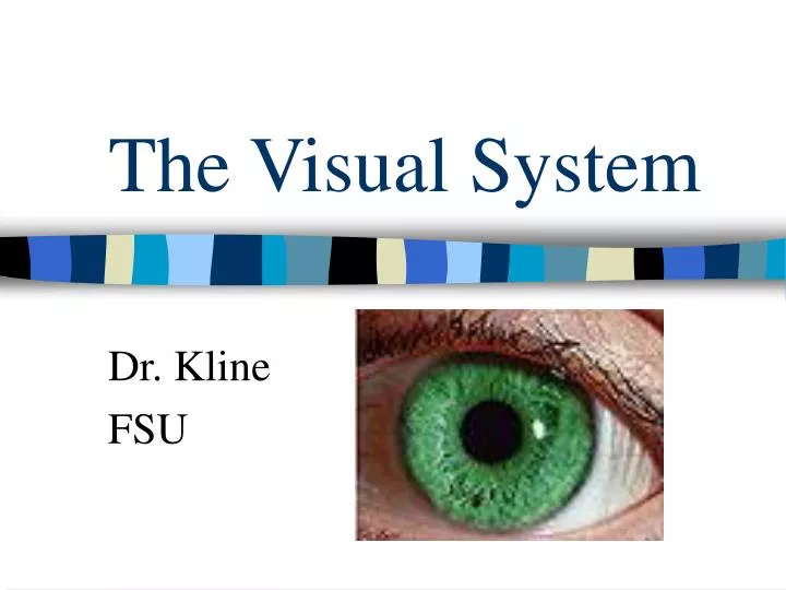
The Visual System. Dr. Kline FSU. What stimuli are required for vision?. Light - which can be thought of as discrete particles (photons) or traveling waves. Human Visible Spectrum. Humans can detect waves of energy traveling through space between 380 & 760 nanometers).

E N D
The Visual System Dr. Kline FSU
What stimuli are required for vision? • Light- which can be thought of as discrete particles (photons) or traveling waves.
Human Visible Spectrum • Humans can detect waves of energy traveling through space between 380 & 760 nanometers). • Wavelengths outside this spectrum are undetectable to the human eye. • Some organisms do detect wavelengths outside our visible spectrum. • E.g., Rattlesnakes detect in infrared!; • bees detect ultraviolet light.
What are the 2 properties of light that influence visual perception? • 1. Wavelength is associated with our perception of color. • 2. Intensity is associated with our perception of “brightness.”
Reflectance of light • Light is reflected off of the surface of objects & to the eye. • Light energy is converted into neural energy & then processed by the brain.
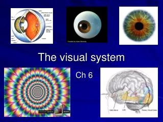
Parts of the eye • Outer Layer • 1. Sclera: White fibrous layer • 2. Cornea: the clear protruding structure in the front of the eye that is curved. • Bends light rays & is responsible for 70-80% of eye’s focusing ability.
Middle Layer • 1. Choroid: vascularized layer that provides nutrition for retinal cells. Is located between the retina & sclera. • 2. Pigment epithileum: a black pigment found between the choroid & retina. • **Traps photons from light to prevent scattering of photons along the retina, which reduces distortion.
Middle layer (contd.) • 3. Iris: smooth ring of muscle with a central opening (pupil). Gives us our eye color! • 4. Pupil: Pupil changes in size depending on intensity of light. -Intense light, small constricted pupils -Dim light, dilated pupils • 5. Lens: focuses light on retina (convex). -lens is round (nearby objects) -lens is flatter for distant objects
Inner layer • 1. Retina: contains receptor cells needed for neural processing of light. A. Fovea: indentation on retina. -fine discrimination; colors & detail. B. periphery: area on either side of fovea of retina. -detection of light • 2. Optic disk: place where axons exit eye forming optic nerve.
The Photoreceptors: Rods & Cones Cones-specialized for color vision & detail (fovea). Rods-sensitive to light (periphery) • 126 million receptors total! • 120 million are rods & 6 million are cones • sensitivity to light enhanced by “convergence ratio.” Many rods converge on a single retinal ganglion cell than do cones. • Rods-big receptive fields • cones-small receptive fields
How does visual information get from the eye to the brain to be processed? • Two vision pathways: • Geniculostriate & Tectopulvinar
Visual Pathways • 1. Geniculostriate pathway- • optic chiasm----LGN---Primary Visual cortex **involved in patter perception, color vision** • 2. Tectopulvinar pathway- • optic chiasm---superior colliculus---Lateral • posterior pulvinar---PVC **detection of light; spatial orientation**
How do our eyes adapt to the dark? • The photopic & scotopic systems adjust their sensitivity as a function of time in the dark. • The cones become more sensitive during the first 5-10 minutes after being in the dark. • Rods continue becoming more sensitive over period of 20-30 minutes.
How is a dark adaptation experiment conducted? • Step 1: The subject (S) is exposed to bright light of a known intensity. • Step 2: S is then placed in total darkness & asked to detect a spot of light (controlled luminance). • Step 3: S’s detection threshold is plotted as a function of time spent in dark.
We have three cone wavelengths • 1. Short wavelength: peaks at 419 nm (blues). • 2. Medium wavelength: peaks at 531 nm (greens). • 3. Long wavelength: peaks at 558 nm (reds). • The primary colors are blue, green, & red
What is relationship between activity in the retina & the brain? • An electrode is inserted into various parts of the visual system (retina, cortex) of an animal. • The cell’s activity in response to the presentation of visual stimuli (lights, bars, complex images) to the animal’s retina is recorded.
What is a receptive field of retinal ganglion cells? • The receptive field for these cells is the region of the retina that, when stimulated excites or inhibits the cell’s firing pattern.
Retinal ganglion cells Kuffler (1953) presented spots of light to retina cells in the cat & recorded their responses. • The cells have a Concentric circle configuration! • usually called center-surround cells • On-center, off-surround cell has an “excitatory center,” & “inhibitory surround” • Off-center, on-surround cell has an “inhibitory center” & “excitatory surround”
Are receptive fields of cortical cells like those of retinal ganglion cells? • No!! • Our visual cortex cells respond to more complex stimuli (e.g., bars of light).
Hubel & Wiesel (1950s & 60s) • Recorded cortical cells in the visual cortex of cats in response to visual images they presented to the cat’s retina. • They found three types of cells with different receptive fields (bar detectors).
Receptive Fields of cortical neurons—Primary Visual cortex • 1. Simple Cells --respond to points of light or bars of light in a particular orientation • 2. Complex cells --respond to bars of light in a particular orientation moving in a specific direction. 3. Hypercomplex Cells: respond to bars of light in a particular orientation, moving in a specific direction, & of a specific line length.
What is the organization of the visual cortex? • Hubel & Wiesel found that the visual cortex is organized into columns. • Location specific: For each place on the retina there is a column of cells in cortex. • Two columns next to one another in the cortex respond to stimulation of two adjacent points on the retina.
Orientation & Ocular Dominance columns in Primary Visual Cortex
The Visual cortex has a retinotopic map • Visual cortex has a map of the retina’s surface. • More cortical neurons are devoted to fovea of retina. • As fovea only has cones, they are widely mapped on cortex’s surface. • The reason: cones allow us to see detail & color.
How do we see in color? • Two Theories: • 1. Young-Helmholtz Trichromatic theory of color vision. The pattern of activity among the three cone-types determines the color we perceive. The cones all respond equally to white light (which contains all wavelengths).
Evidence for Trichromatic theory • 1. We have three types of cones sensitive to different wavelengths of light (short, medium, & long wavelengths). • 2. There are 3 types of color-blindness. • 3. Approximately any color can be matched by mixing varying amounts of red, green, & blue light.
Color-blindness • Results whenever we are either missing one of our cones or one of our cones doesn’t work properly.
2. Opponent-Process theory • We have opposing mechanisms that allow us to perceive colors. Evidence for this theory: 1. Incompatible colors cannot be seen Explains why we can’t see certain colors (reddish-green, bluish-yellow) & color afterimages. We have 3 opposing mechanisms: red-green, yellow-blue, & black-white. These are called complimentary colors & put together they produce yellow or white.