Download
1 / 105
1.07k likes | 1.31k Views
Refresher Training for X-Ray Equipment Operators . Developed for the Department of Environmental Protection Bureau of Radiation Protection. Presented by David R. Simpson, CHP, PhD Bloomsburg University. Outline of Course. Part 1: Introduction and Overview of the Training Program

E N D
Refresher Training for X-Ray Equipment Operators Developed for the Department of Environmental Protection Bureau of Radiation Protection Presented by David R. Simpson, CHP, PhD Bloomsburg University
Outline of Course • Part 1: Introduction and Overview of the Training Program • Part 2: Fundamentals of Radiation Science • Part 3: Review of X-ray Imaging • Part 4: Safety Plan, Documentation and QA • Part 5: Regulations
Part 1 Introduction and Overview of Training Program
Introduction and Background • X-rays a key component of diagnostic medicine for many years • BUT will result in exposure of patient and staff to ionizing radiation • Food and Drug Administration (FDA) recommends two steps for medical X-rays: • Justification of procedure • Optimization of procedure
Justification of Imaging Procedure • Imaging procedure should do more good than harm; therefore, exams should be performed only when necessary to: • Answer a medical question • Treat a disease • Guide a procedure
Optimization of Imaging Procedure • Exams should use techniques that are adjusted to: • Administer lowest radiation dose that yields image quality adequate for diagnosis or intervention • That is, radiation doses should be “As Low As Reasonably Achievable,” referred to as the ALARA Principle
Purpose of Training • According to the National Council on Radiation Protection and Measurements (NCRP Report No. 134) there are four major reasons for training: 1. Development of worker skills so that tasks may be performed efficiently and with confidence
Purpose of Training 2. Individuals aware of risk of exposure become active participants in accepting and, where possible, reducing those risks 3. Number and seriousness of accidents can be reduced 4. Workers will be aware of regulatory requirements involved with radiation exposure
State Requirements for Training • Pennsylvania Department of Environmental Protection (DEP) regulations require individuals operating X-ray equipment to: • Receive initial instructions in safe operating procedures • Be competent in the safe use of equipment • Receive continuing education
State Requirements for Continuing Education DEP Technical Guidance Document No. 291-4200-001 lists the following topics for continuing education: • Basic properties of radiation • Units of measurement • Sources of radiation exposure • Methods of radiation protection • Biological effects of radiation exposure
Topics for Continuing Education • X-ray equipment • Image recording and processing • Patient exposure and positioning • Procedures • Quality assurance program • Regulations
To provide the generic portions of this training for operators performing low-risk medical procedures Training Goal
Part 2 Fundamentals of Radiation Science
Fundamental Properties of Radiation for X-ray Imaging • Properties of Radiation • Units of Measurement • Sources of Radiation • Biological Effects of Radiation
Ionizing Radiation • High energy particles or electromagnetic (EM) energy • Capable of removing orbital electrons from atoms • Effect called ionization • Resulting atom and electron called ion pair
Particulate vs. EM Ionizing Radiation • Particulate • Includes alpha, beta, neutrons • Usually easier to shield • Not used in diagnostic medicine (some applications in therapy) • EM • Includes gamma, and X-rays • Generally more difficult to shield • X-rays are major tool in diagnostic medicine
Types of EM Radiation • Most EM radiation is non-ionizing • Common names for various energies of EM radiation: • Radio waves (very low energy, non-ionizing) • Microwaves (non-ionizing despite common perception) • Infra-red, visible, and Ultraviolet (non-ionizing, though intense forms may damage skin or eyes) • X-rays (ionizing) • Gamma rays and cosmic rays (ionizing)
X-rays vs. Other Forms of Ionizing Radiation • X-rays (with some minor exceptions) are produced by machines. • Particulate radiations and gamma rays primarily come from radioactive materials. • X-rays cannot make a person radioactive and cannot result in contamination (loose radioactive materials).
X-rays vs. Other Forms of Ionizing Radiation X-ray machines have an on-off switch They can be immediately turned off, removing the source of radiation
Quantities & Their Units of Measurement • Three Basic Quantities • Exposure – measure of charge in air produced by X-ray or gamma radiation • Absorbed Dose – measure of energy deposited by any type of ionizing radiation in any material • Equivalent Dose – measure of biological damage to the human caused by various types of ionizing radiation
Why Three Quantities? • Exposure • Easy to measure using inexpensive instruments • Can be related to other two quantities • Absorbed Dose • Widely applicable to measuring effects of radiation • Often difficult to measure • Equivalent Dose (or Dose equivalent) • Allows for biological differences in humans for different types of radiations • Used for regulatory limits
Exposure Units • Roentgen • Oldest unit, defined as 0.000258 coulombs/kg in air • Still widely used in USA but not defined in the current international scientific unit system (SI) • Symbol: R • Air kerma (closest SI equivalent) • Defined as 1 joule/kg in air • Symbol: Gya (grays in air – see absorbed dose) • To a close approximation 1R = 0.01 Gya
Absorbed Dose Units • rad • Traditional unit defined as 100 ergs/g of energy deposited by any type of ionizing radiation in any mass (g) of material • Gray (Gy) • SI unit defined as 1 joule/kg of energy deposited by any type of ionizing radiation in any mass (kg) of material • 1 rad = 0.01 Gy
Equivalent Dose Units • rem – traditional unit defined as absorbed dose in rad multiplied by modifying factors to account for responses in the human • Sievert (Sv) – SI unit defined as absorbed dose in gray multiplied by modifying factors to account for responses in the human • 1 rem = 0.01 Sv
Relationship of Units • For X-ray radiation, to a close approximation we can assume that: • 1 R = 1 rad = 1 rem and • 1 Gya= 1 Gy = 1 Sv
Dose and Dose Rate • Radiation often measured as a dose rate (dose per time) so dose received is calculated as follows: • Dose received = Dose Rate x Time exposed
Example of Dose Rate & Conversions Exposure rate in area is 2 mR/hr. Is yearly limit of 5 rem exceeded? (Assume occupancy of 40 hours/week and 50 work weeks in year). • Exposure = 2mR/hr x 40 hr/wk x 50 wks/yr • Exposure = 4000 mR/yr = 4 R/yr • Assume 1R = 1 rad = 1 rem, so • Equivalent dose = 4 rem/yr; limit not exceeded
Sources of Radiation • Radiation exposure to the general public comes from a number of sources: • Natural background radiation (in soil, air, our bodies, etc.) • Medical procedures • Occupational exposures • Consumer products • Following slide shows breakdown of exposures and recent trends
So what has happened? • Background levels are the same, but in % they have decreased to only about half of the total average exposure • Occupational and consumer product levels have remained very low • Medical exposures have increased significantly (on the average) • Why?
So what has changed? • Large increase in number of CT scans • Increase in nuclear medicine procedures • Newer techniques involve higher doses to the patient
High-Risk vs. Low-Risk Procedures Pennsylvania has divided X-ray medical procedures into two risk classes: • High-risk procedures – utilize energies of < 1 MeV that could exceed skin doses of 200 rads (2 Gy). • Examples: CT scans, interventional radiography • Low-risk procedures – any radiologic procedure that is not a “high-risk” procedure. • Examples: conventional x-rays, dental, podiatry, chiropractic, and veterinary High-risk results in more dose per patient, but many more patients receive low-risk procedures (especially children and young adults).
Biological Effects of Radiation • Harmful effects discovered very early • (Thomas Edison ceased work on X-rays in 1904 following a serious injury and subsequent death of his assistant due to radiation) • “Law of Bergonie and Tribondeau” developed in France in 1906: • “The more rapidly a cell is dividing, the greater the sensitivity to radiation” • Not always true but helpful in explaining effects on certain organs such as skin, blood-forming organs, gonads and unborn children.
What is the primary biological effect? • Research has found that the primary hazard is to DNA • Can result in cell death leading to organ failure, illness, and possible death of exposed person • Or can result in cell mutations leading to cancer in exposed person and possible genetic effects to future generations • Greatest risk at low doses is cancer to exposed person – regulations based on this risk
Acute vs. Delayed Effects • Acute effects (also referred to as early effects or deterministic effects) • Typically occur at high doses and appear within days or weeks of exposure • Examples: skin burns, sterility, loss of hair, etc. • Delayed effects (also referred to as late effects or stochastic effects) • Occur at low doses over long periods of time • Cancer is greatest concern
Goal of Radiation Regulations • Prevent acute effects to exposed person • Shield sensitive organs such as lens of eye, thyroid, unborn child • Reduce likelihood of delayed effects • Keep doses low and make sure they are justified • In other words, ALARA: As low as reasonably achievable
Part 3 Review of X-ray Imaging
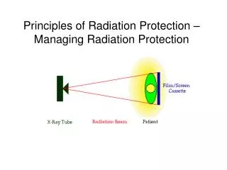
Basic Components of X-ray Tube • Cathode – heated wire to produce large source of electrons and focusing cup to direct them • Anode – target of high atomic number struck by electrons to produce X-rays • Voltage supply – high voltage supply to accelerate electrons from cathode to anode • Envelope – glass or metal vacuum tube containing anode and cathode • Tube housing – shielding around envelope to protect tube and shield unwanted X-rays
Other Parts of X-ray Machine • Collimator – restricts X-ray beam to only the area of interest • Filtration – removes unwanted low energy X-rays • Transformer – converts low voltage to high voltage needed for tube • Rectifier – converts AC input voltage to DC needed for X-ray tube
Primary Settings on X-ray Tube • Current to cathode – measured in milliamps (mA) • Timer for current in seconds • Current and timer together are measured in mAs • Voltage across the cathode and anode – measured in kVp (kilovoltage peak, the maximum possible energy a photon exiting the X-ray tube can reach) • kVp determines the energy of electrons which is directly related to energy of X-rays produced
Quantity and Quality of X-rays • Medical X-rays characterized by quantity and quality • Quantity – number of X-rays reaching the patient • Quality – penetrability or ability of X-ray beam to pass through tissue (low quality X-rays have little chance of penetrating so they deliver dose to patient while providing no useful medical information)
Impact of Digital Imaging • Digital (computer) imaging replacing conventional (film) systems • Images immediately available, can be stored and transmitted electronically and post processed to improve image after the fact • Should result in lower dose to patients due to fewer retakes
Dose Creep in Digital Imaging • In some cases doses increased in digital systems • Effect called dose creep • Digital resulted in good images without changing factors, even if patient dose was higher, so factors were not optimized to lower dose. • Also, techniques were used to reduce signal noise that resulted in increased dose. • Manufacturer’s recommendations should be reviewed to minimize dose to patient
Reducing Patient (and employee) Dose from Medical X-rays • Sources of radiation in Medical X-ray Imaging: • Primary beam – also called the useful beam; the X-ray beam coming from the tube, through the patient, to the image receptor. • Scatter radiation – radiation resulting from the primary beam interacting with other materials. The patient is often the largest source of scatter. • Leakage radiation – leakage radiation from tube housing, generally a minor source from a properly housed tube.