Download
1 / 24
270 likes | 782 Views
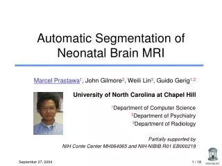
Automatic Segmentation of Neonatal Brain MRI. Marcel Prastawa 1 , John Gilmore 2 , Weili Lin 3 , Guido Gerig 1,2 University of North Carolina at Chapel Hill 1 Department of Computer Science 2 Department of Psychiatry 3 Department of Radiology Partially supported by

E N D
Automatic Segmentation of Neonatal Brain MRI Marcel Prastawa1, John Gilmore2, Weili Lin3, Guido Gerig1,2 University of North Carolina at Chapel Hill 1Department of Computer Science 2Department of Psychiatry 3Department of Radiology Partially supported by NIH Conte Center MH064065 and NIH-NIBIB R01 EB000219
Goal • Segmentation of brain tissues of newborn infants from multimodal MRI • Particular interest in the developing white matter structure Motivation: Analysis of growth patterns, study of neuro-developmental disorders starting at a very early age csf nWM gm mWM Automatic Segmentation of Neonatal Brain MRI
Imaging the Developing Brain age 35 weeks 44 weeks 15 months 2 years adult Rhesus equiv. 6yrs Automatic Segmentation of Neonatal Brain MRI
T2 T1 Challenges • Smaller head size • Low contrast-to-noise ratio • Intensity inhomogeneity • Motion artifacts • Division of white matter into myelinated and non-myelinated regions Previous work: Warfield et al 1998 (methodology) Hüppi et al 1998 (clinical study) T1 T2 Labels csf nonm. WM gm myel. WM Automatic Segmentation of Neonatal Brain MRI
Challenges Neonate 2 weeks Adult CNR 2.9 CNR 6.9 Automatic Segmentation of Neonatal Brain MRI
Approach • Non-optimal input data, rely on high level prior knowledge • Intensity ordering (e.g. in T2W) wm-myelinated < gm < wm-non-myelinated < csf • Aligned spatial priors (brain atlas) White matter is considered as one entity Automatic Segmentation of Neonatal Brain MRI
Compute posteriors Intensity Clustering Whole brain MST clustering Bias correction Compute PDFs Parzen Windowing Method Overview Segmentation - Bias correction Initialization Refinement Automatic Segmentation of Neonatal Brain MRI
Intensity Clustering • Samples obtained by thresholding atlas priors T1 T2 Pr(wm, x) Overlay • Noisy data, low contrast robust techniques • Two robust estimation techniques: • Minimum Spanning Tree (MST) clustering • Minimum Covariance Determinant (MCD) estimator • Obtain initial estimates of intensity distributions Automatic Segmentation of Neonatal Brain MRI
Minimum Spanning Tree Clustering [Cocosco et al 2003] • Break long edges in MST, example: Detect multiple clusters while pruning outliers • Iterative process, stops when cluster feature locations are in the desired order • “Feature location” = summary value of cluster intensities Automatic Segmentation of Neonatal Brain MRI
Determining Feature Locations • Need reliable location estimate to find good clusters • Standard estimates (e.g., mean, median) not always optimal • Use robust estimator to determine location of a compact point set in a cluster Median Mean Automatic Segmentation of Neonatal Brain MRI
O = points used for estimation X = other data points Minimum Covariance Determinant [Rousseeuw et al 1999] • Feature location of MST clusters to determine ordering? • Smallest ellipsoid that covers at least half the data • MCD gives robust location estimate • Example: Automatic Segmentation of Neonatal Brain MRI
csf nWM gm mWM T2 T1 Intensity Clustering Algorithm Apply MCD to GM and CSF samples: obtain T2 locations Construct MST from WM samples T 2 Repeat until T = 1 Break edges longer than T x (local average length) Find largest myelinated WM cluster, where: T2myel< T2GM Find largest non-myelin. WM cluster, where: T2GM < T2non-myel< T2CSF Stop if WM clusters found Otherwise, T T – 0.01 Automatic Segmentation of Neonatal Brain MRI
Compute posteriors Intensity Clustering Whole brain MST clustering Bias correction Compute PDFs Parzen Windowing Method Overview Segmentation - Bias correction Initialization Refinement Initial intensity Gaussian PDFs Automatic Segmentation of Neonatal Brain MRI
biology bias Compute posteriors Bias correction Compute PDFs Bias Correction [Wells et al 1996, van Leemput et al 1999] • “Bias” = RF inhomogeneity and biology • Images low contrast, histogram is smooth • Use spatial context, bias is log-difference of input intensities and reconstructed “flat” image • Fit polynomial to the bias field (weighted least squares) • Interleaves segmentation and bias correction Gaussian intensity PDFs Automatic Segmentation of Neonatal Brain MRI
Compute posteriors Intensity Clustering Whole brain MST clustering Bias correction Compute PDFs Parzen Windowing Method Overview Segmentation - Bias correction Initialization Refinement Bias corrected images Segmentations Automatic Segmentation of Neonatal Brain MRI
Refinement • Previous stage assumes Gaussian intensity distributions • May have non-optimal decision boundaries due to overlap • Re-estimate intensity parameters from bias-corrected images • MST clustering to obtain training data • Parzen windowing to estimate density Parzen kernel density estimate Atlas prior Automatic Segmentation of Neonatal Brain MRI
Results [1/2] • UNC Radiology Weili Lin (Siemens 3T head-only) • UNC-0094 • UNC-0096 Classification T1 T2 3D Classification T1 T2 3D Automatic Segmentation of Neonatal Brain MRI
Results [2/2] • Provided by Petra Hüppi (Geneva, Philips 1.5T) • Geneva-001 • Geneva-002 T2 Classification T1 3D T2 Classification T1 3D Automatic Segmentation of Neonatal Brain MRI
Results: UNC 0096 Upper row: T1, T2w, Tissue labels, registered atlas Lower row: Probabilities for wm-myel, wm, gm, csf Automatic Segmentation of Neonatal Brain MRI
Results: UNC 0096 Automatic Segmentation of Neonatal Brain MRI
Summary • Automatic brain tissue segmentation of neonatal MRI • Detects white matter as myelinated and non-myelinated structures • Makes use of prior knowledge: • Image intensity ordering • Spatial locations (probabilistic atlas prior) • To be used in two large UNC neonatal MRI studies • Silvio Conte Center: 125 neonates at risk • Neonate Twin study (heritability) • Current focus: Validation Automatic Segmentation of Neonatal Brain MRI
Acknowledgements • Elizabeth Bullitt • Petra Hüppi • Koen van Leemput • Insight Toolkit Community Neoseg v1.0b Automatic Segmentation of Neonatal Brain MRI
Validation (in progress) A) Semiautomated expert segmentation of a few cases • Edge-based segmentation • Level-set evolution • Manual editing • Primarily: White-gray contour B) Simulated MRI data (similar to MNI ICBM) Automatic Segmentation of Neonatal Brain MRI
New Probabilistic Atlas for the 2yrs group 14 subjects, aligned, intensity adjusted, segmented (UNC M. Jomier/Piven/Cody/Gimpel/Gerig) Automatic Segmentation of Neonatal Brain MRI