Download
1 / 23
240 likes | 523 Views
Cardiac Cycle and Heart Sounds. Conduction system of the heart. a. Sinoatrial node (SA node ) b. Atrioventricular node (AV node) c. Atrioventricular bundle (AV bundle) d. Right and left atrioventricular bundle branches e. Purkinje fibers. Sinoatrial Node.

E N D
Conduction system of the heart • a. Sinoatrial node (SA node) • b. Atrioventricular node (AV node) • c. Atrioventricular bundle (AV bundle) • d. Right and left atrioventricular bundle branches • e. Purkinje fibers
Sinoatrial Node • Located Junction of Superior Vena Cava • Specialized Pacemaker Cells • Intrinsic Rhythm- without stimulation by nerve impulses from brain and spinal cord • Initiates impulses at regular intervals
AtrioVentricular Nodes • From SA node to Contraction of both Atriums • Internodal Bundles- Right Atrium • Interartial Bundle- Left Atrium • Internodal Bundles to Atrioventricular node
Bundle of His • Av Node to Bundle of His (AV Bundle) • Left and Right Bundle Branches • Branches to Purkinje Fibers • Simultaneously Contract Ventricles
HeartBEat • SA node Intrinsic Rhythmical rate 70-75 beats • What happens when SA node loses ability to initiate impulse? • Another Excitable Component takes over • Abnormal/ ectopic pacemakers • AV Node or Purkinje Fibers • Slower rate than SA Node • AV Node- 40-60 beats per min
Conduction Of the Heart Video • Heart Conduction Animation • McGraw Hill
ElectroCardiogram (ECG) • ECG (EKG) • a recording of the electrical activity (changes) during a cardiac cycle • How it works • Two Electrodes of Voltmeter • Passing of Action Potential between two electrodes
ECG Waves • P Wave – depolarization(+) of the atria (atrial contraction – systole) • SA Node Atria • QRS Complex – depolarization of the ventricles (ventricular contraction, systole) • AV Purkinje Fibers • T Wave – Repolarization(-) of the ventricles
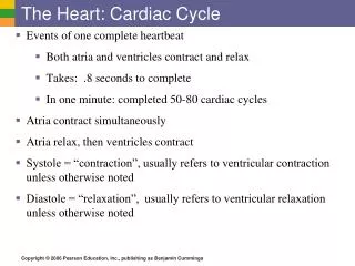
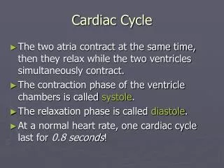
Cardiac Cycle • Cardiac Cycle is all the events associated with the blood flow through the heart during one complete heartbeat • Systole – contraction period of a chamber – ejection of blood. • Diastole – relaxation period of a chamber – filling of blood • Contraction (Eject) and Relaxation (Filling) ALWAYS follows electrical events seen in an ECG
Cardiac Cycle • SA Node initiates action potential. • Atrial systole (1) causes Atrialpressure to increase, blood sent to ventricles through AV valve. (P Wave) • Ventricular Systole caused by impulsetraveling down Bundle of His to PurkinjeFibers (QRS Wave)
Cardiac Cycle • Isovolumetric Ventricular Contraction (2) • AV valves snap close causing the “Lubb” sound at the pressure rises in the ventricles • Ejection (3) of blood into pulmonary circuit • pressure in ventricle is greater than in the arteries so semilunar valves are forced open. • As pressure drops the Semilunar Valves snap close causing “Dubb” sound
Cardiac Cycle • Isovolumetric Ventricular Relaxation (4) • Diastole begins, all valves closed. Once pressure has dropped low enough, the AV valves open and….. • Passive Ventricular Filling (5) because atrial pressure is higher than ventricle pressure. Toward the end of this period, atrial systole begins.
Heart Sounds • Closing of the heart valves • Described as “Lubb – Dubb”Basic rhythm is lubb-dubb, pause, lubb-dubb, pause, lubb-dubb, pause. • First sound “Lubb” is the longest and loudest occurring when AV valves close. • Second sound “Dubb” is short, sharp sound occurring when Semilunar valves close.
Abnormal Sounds – Heart Murmurs • Sound is due to turbulence • Generally, heart murmurs indicate heart valve problem • “Swishing” sound indicates an incompetent valve - partially open valve after it should be closed so the blood backflows through the valve • High-pitched or click sound indicates a stenotic valve – the valve should be wide open, but it is not.